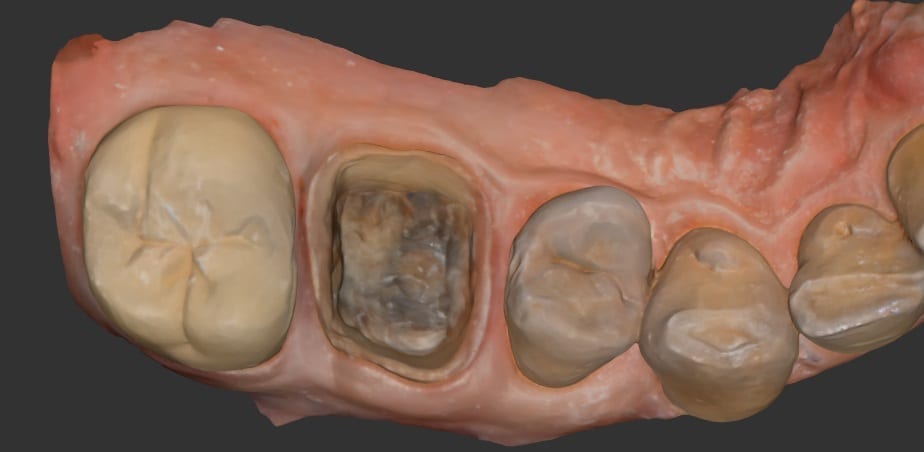
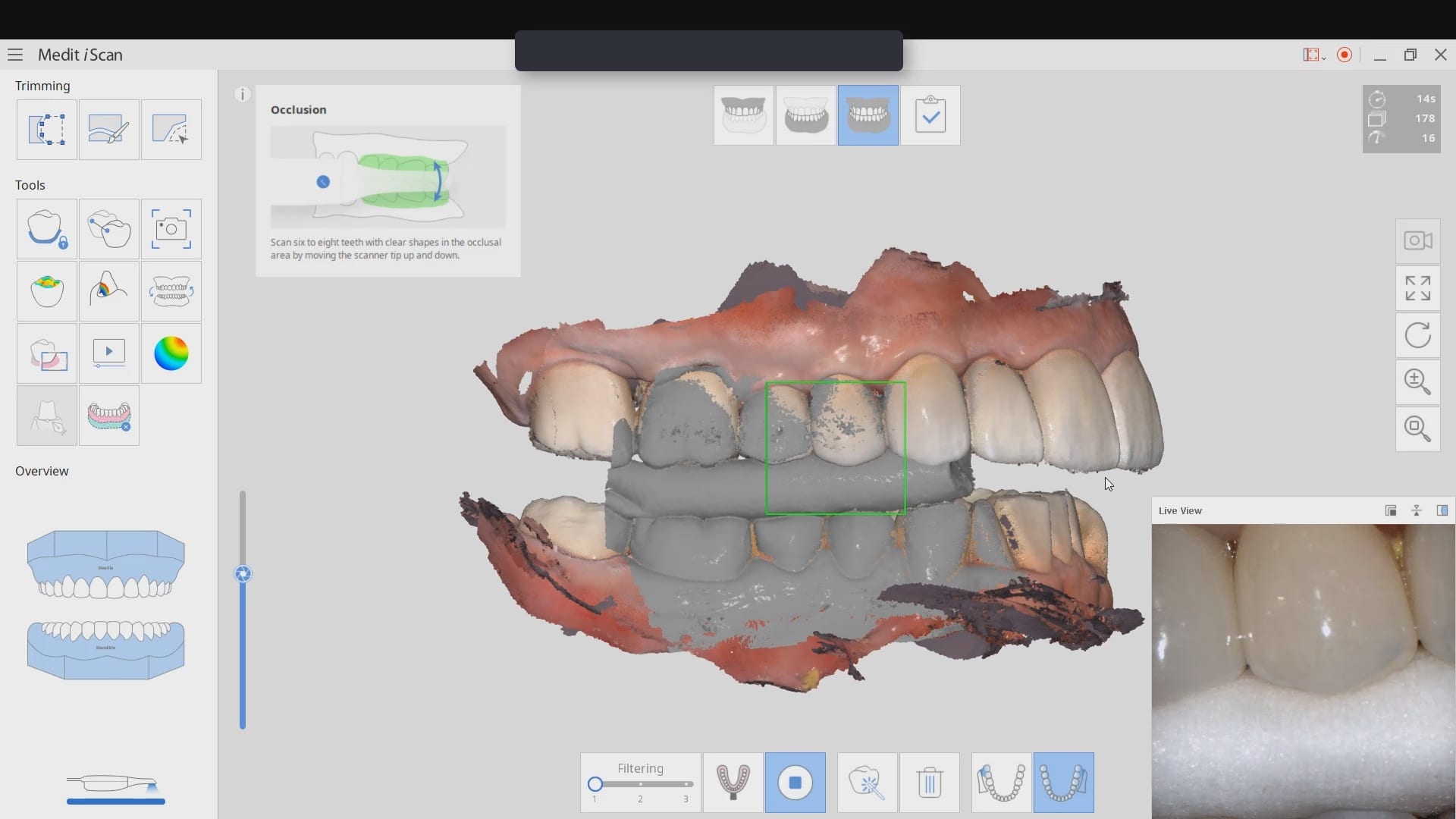
In this video, you can see how easy it is to image both arches and capture all the margins and contacts. There are certain areas that are not necessary for you to capture. For example, in contact areas of adjacent teeth, you do not need to image anything below the height of contour of the adjacent teeth, since you won’t be making contacts to those areas.
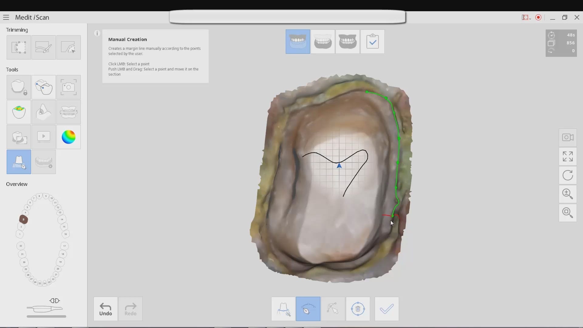
This is an upper and lower case that is scanned with the Medit i500 intra-oral scanner. The lower arch was prepared first and imaged and saved. Then the upper molar was prepared and scanned as well. The bite was captured from the buccal while the patient was biting down in maximum intercuspation. Tissue retraction was not necessary in either case as we could discern the margins simply by the color difference between the tooth structure and the tissue.
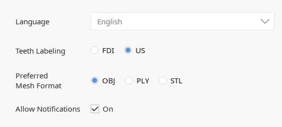 The software allows you to export the file in STL, PLY, or OBJ formats. When just submitting an STL file to the lab, the color difference in the model is not transmitted. But with other formats, you can easily convey to the lab what you see clinically.
Image of case exported as STL
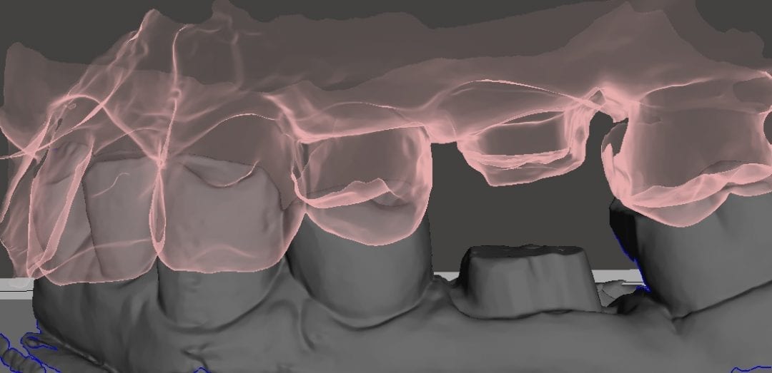
Image of case exported as OBJ
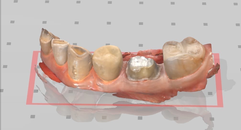
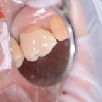
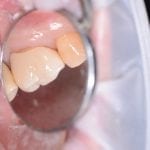
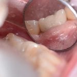
Delivery of the restorations, fabricated by Burbank Dental Lab. There were no adjustments made to the contacts or to the occlusion.





You must log in to post a comment.