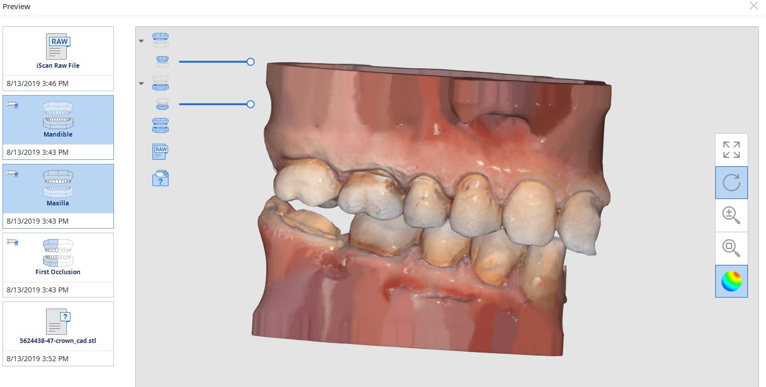
Few things in dentistry that can be as frustrating as seating a second molar restoration, whether you are doing same day dentistry or having a lab made prosthesis delivered. Here is a protocol we recommend that you follow to dramatically reduce surprises and post op adjustments. In this particular clinical case a zirconia crown debonded and we elected to fabricate an in-office emax restoration. The sequence is as follows:
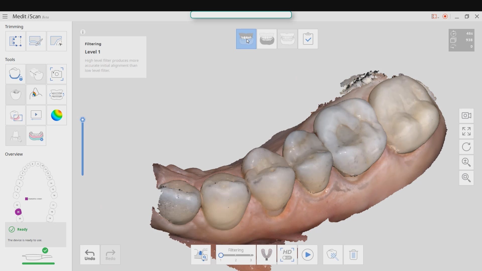
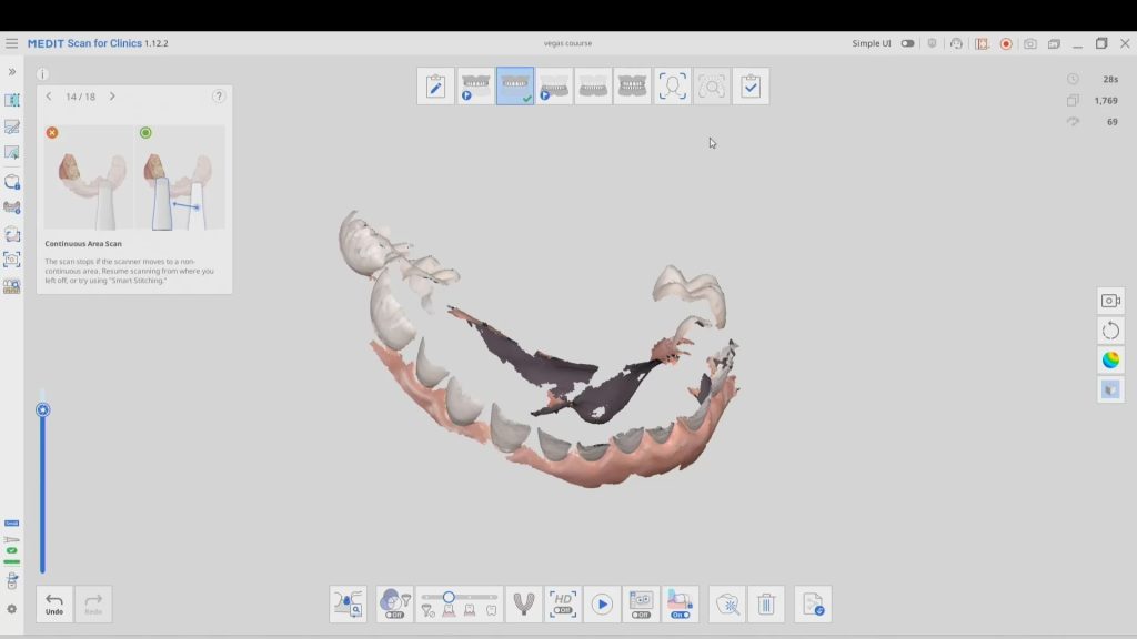
- While the patient is anesthetized and you are waiting for the onset of anesthesia, capture the opposing impression and the arch models. Trim away the prep digitally and then proceed to the buccal bite capture
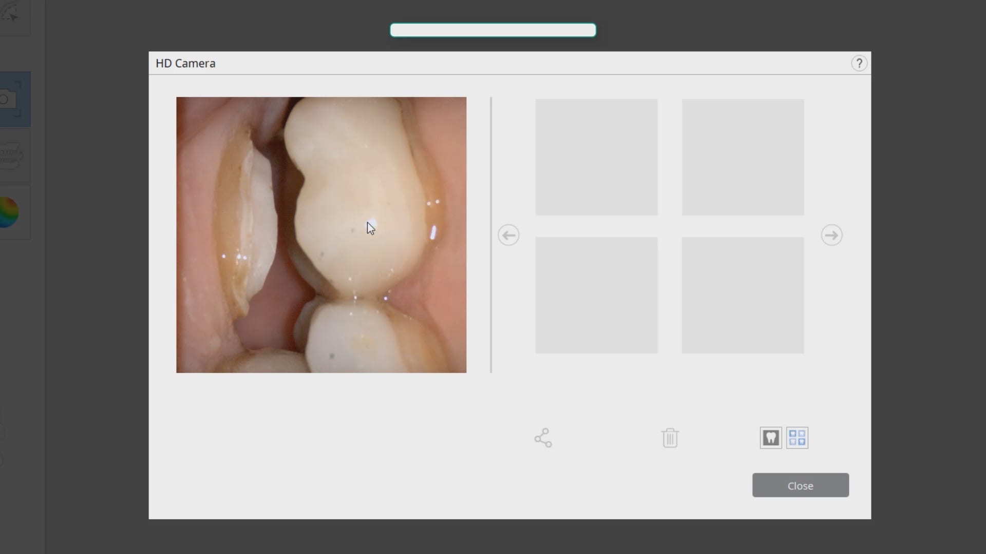
- Do NOT capture the bite until you verify clearance. In the sequences of videos that follow, watch how we use the Medit i500 to capture digital pictures of the clearance
- Once we verify clearance, we image the bite. You have the option at this point to see how well your occlusal stamps match the digital stamps if you want to. A large deviation may mean the jaw settled or the patient moved during the bite capture. Note that unlike conventional dentistry, you capture the bite here BEFORE the prep is finalized
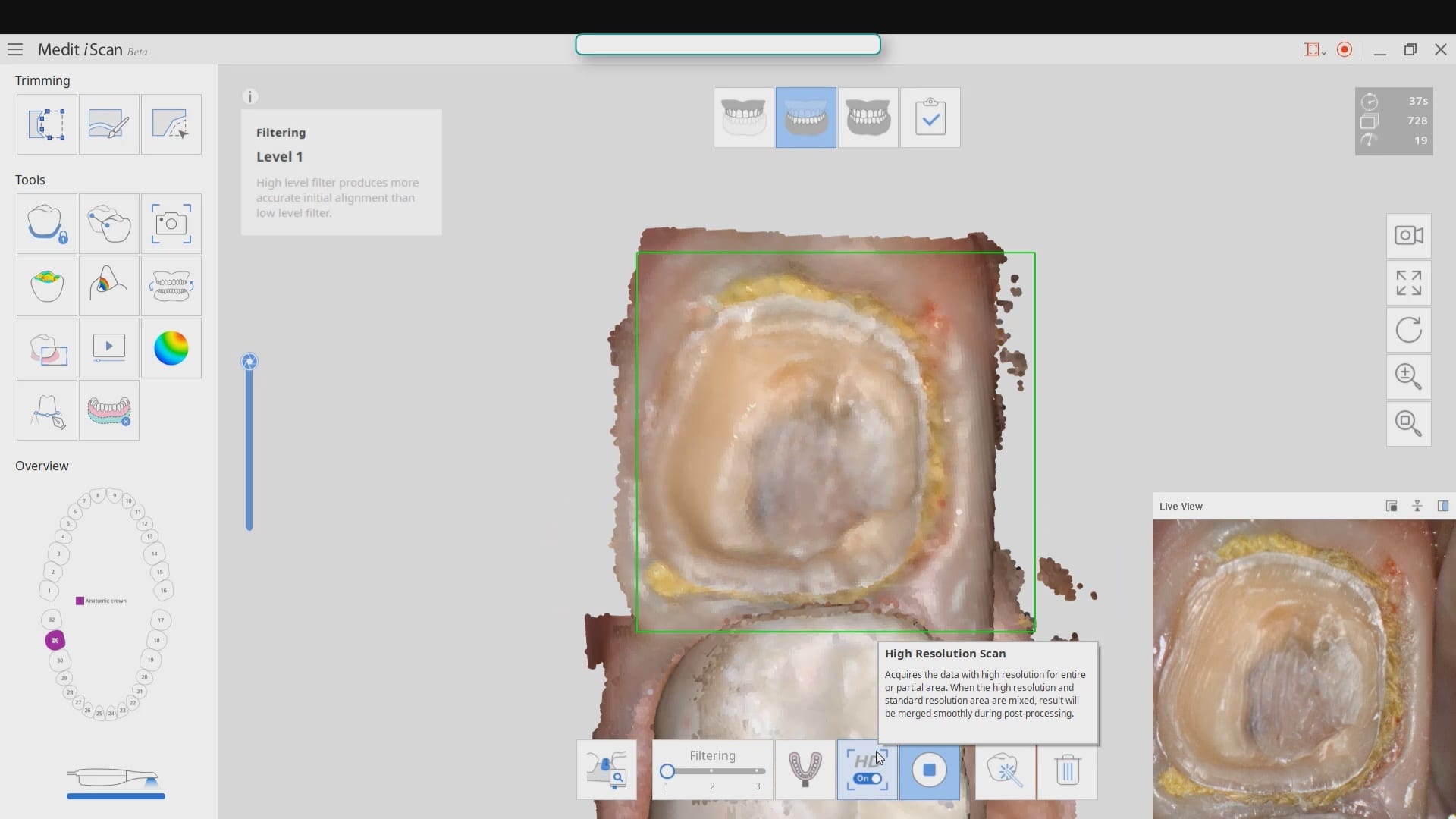
- Once you achieve isolation you can finalize the prep and retract the tissue and capture the prep. We elected to capture the preparation in HD mode
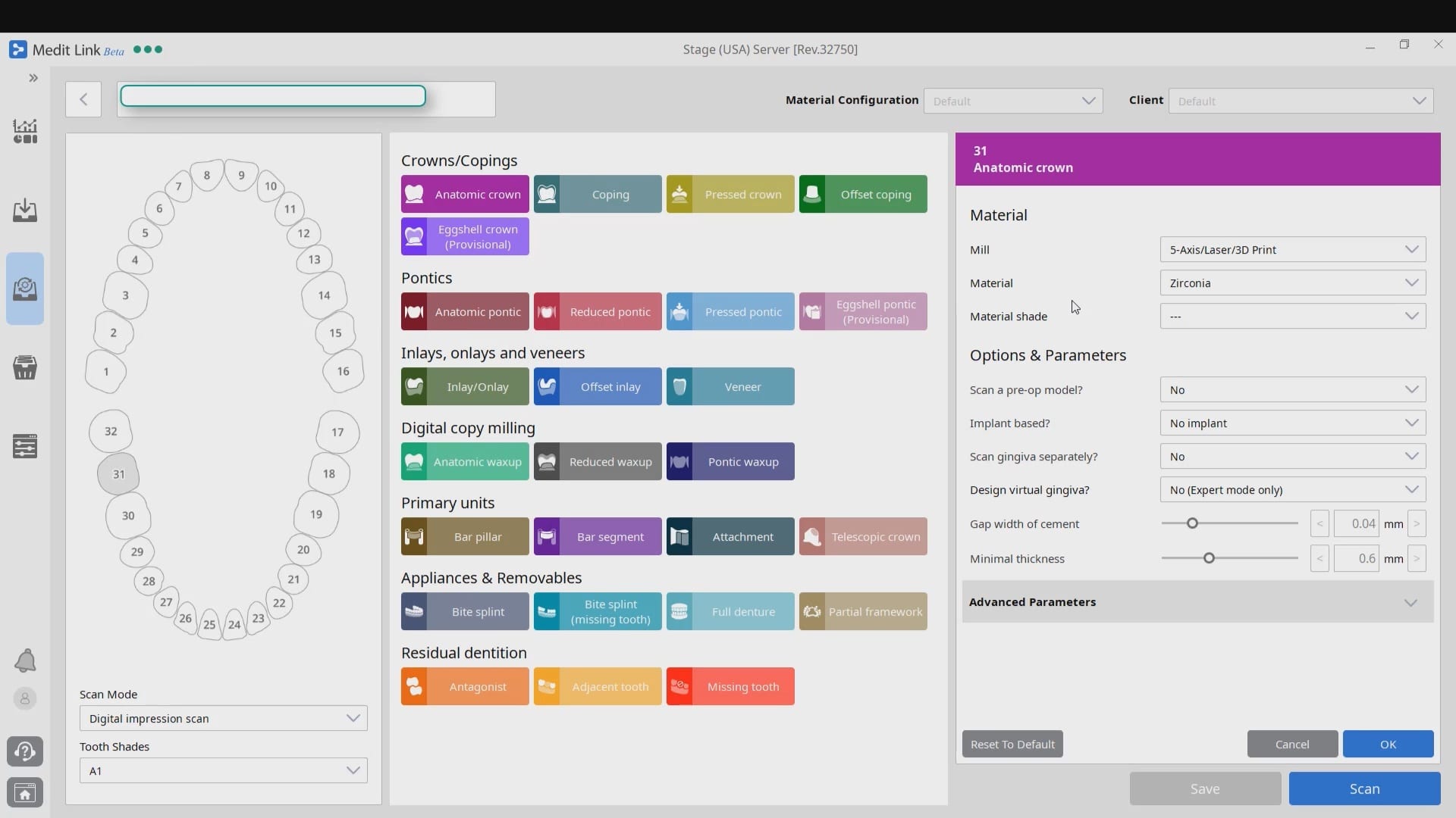
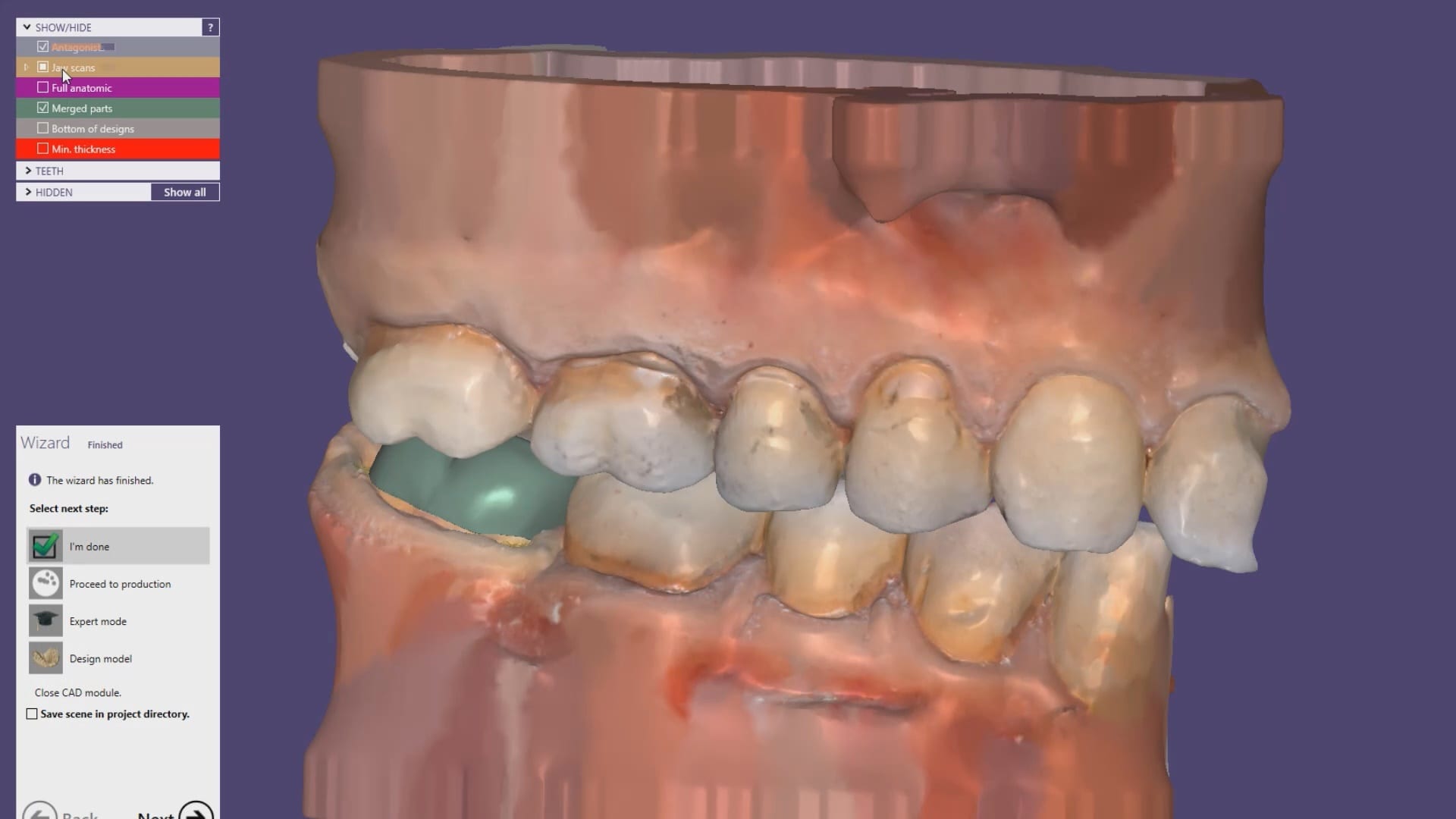
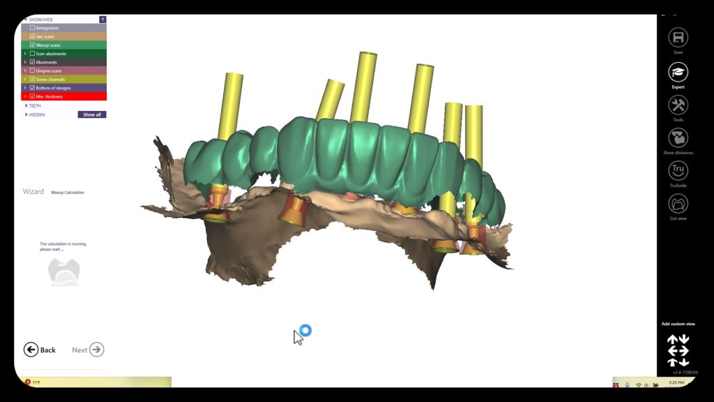
- The case is then immediately imported into the CAD software for design and fabrication
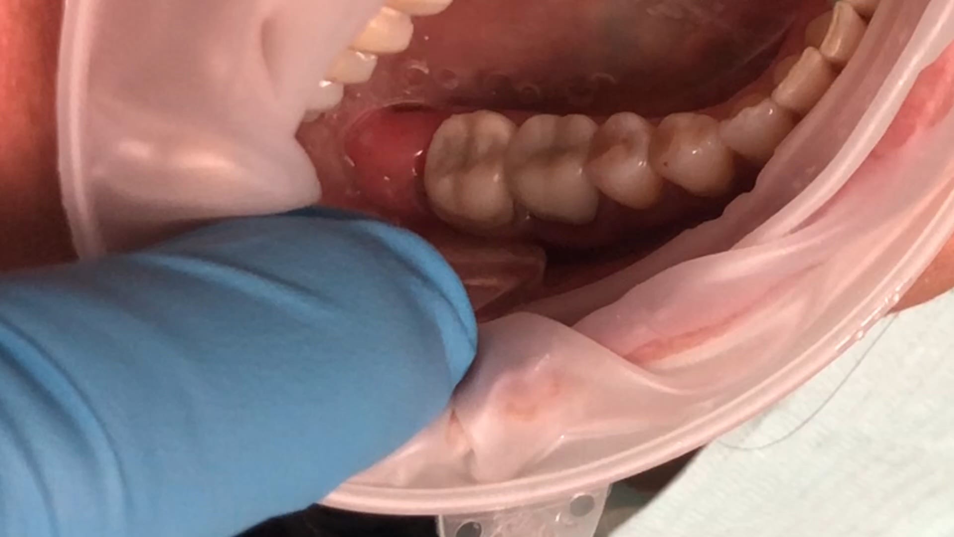
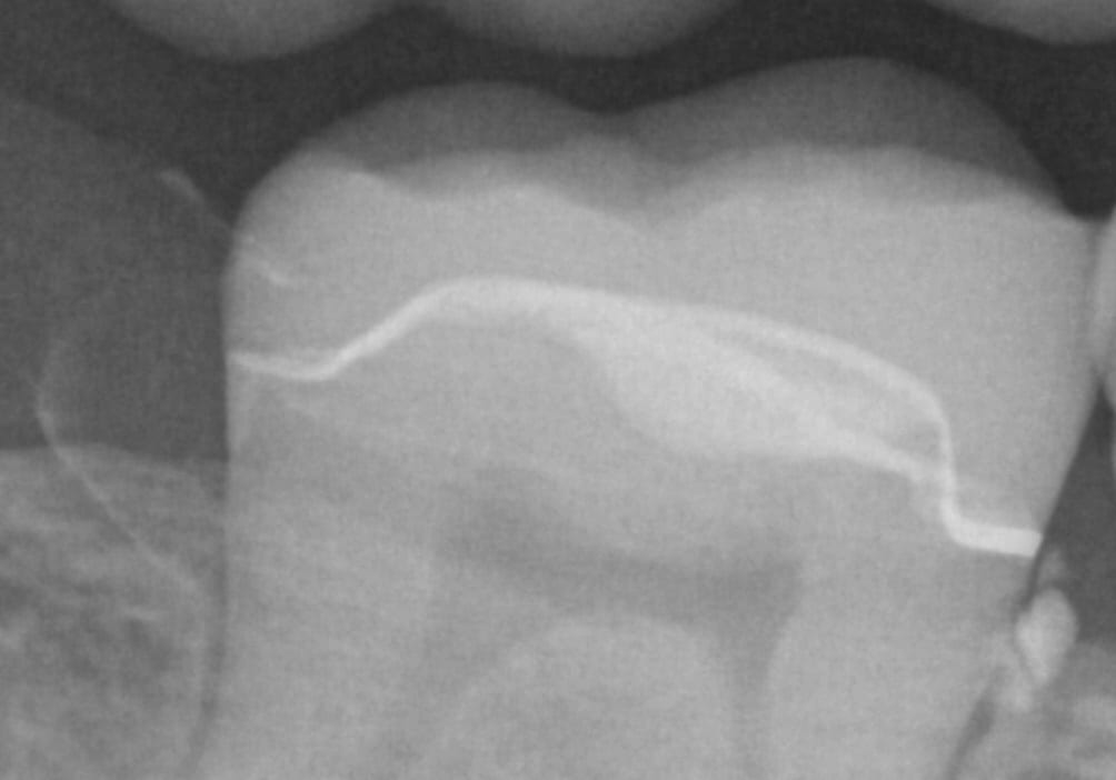
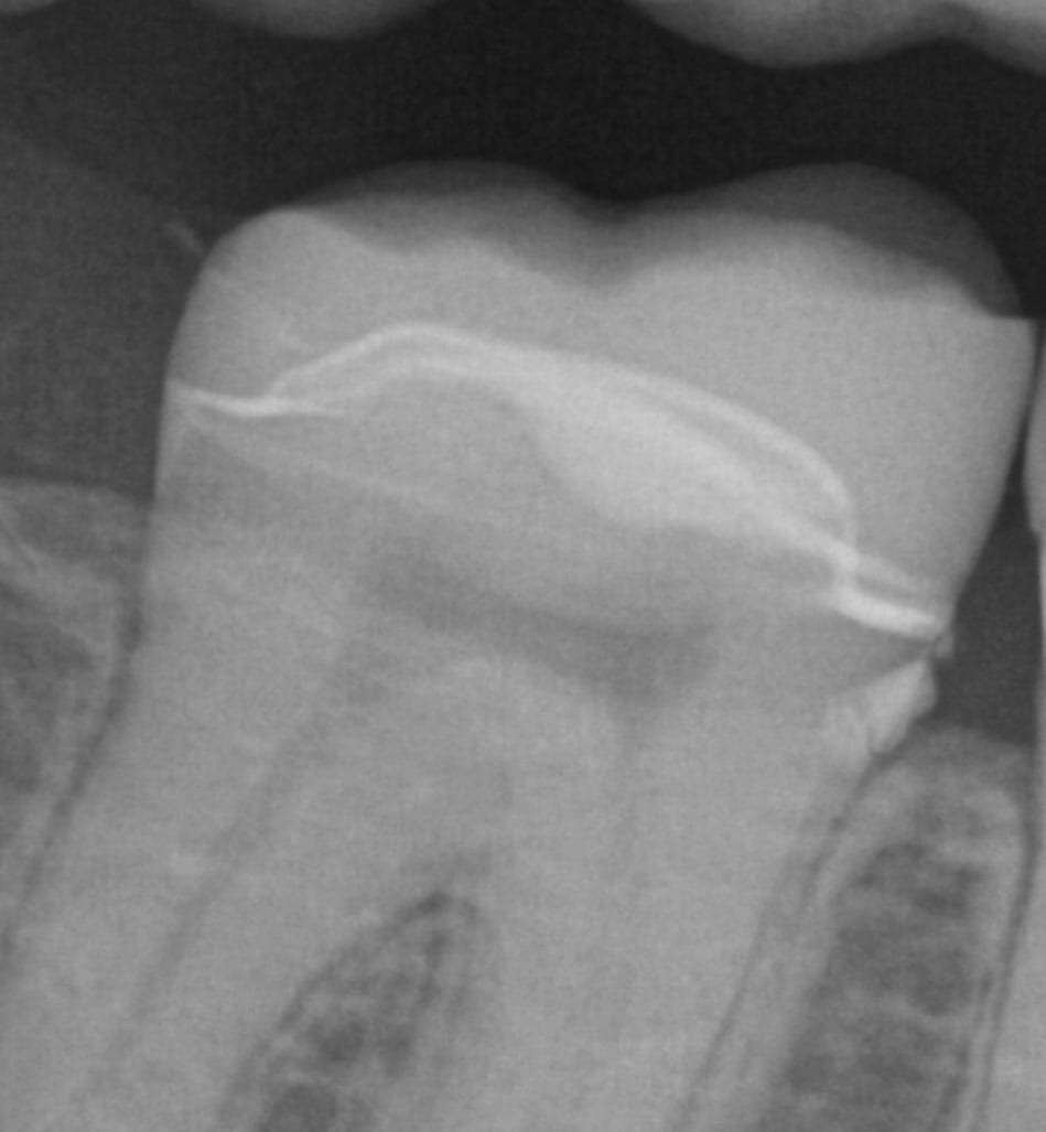
Immediate post op x-rays were taken to verify seat and aid in resin cement removal. The excess cement was removed after the x-rays were taken




You must log in to post a comment.