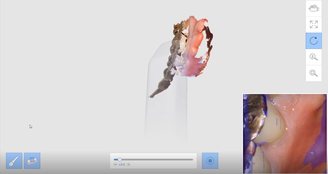
Imaging highly reflective surfaces can be challenging. In this particular situation, every tooth had either a pfm or a gold crown restoration. The video playback feature of the medit software allows you to see exactly how these areas are managed and captured. in the live preview box in the lower right corner, you can see the purple silhouette which shows areas that cannot be seen. Just angle the camera and moving its focal length and angulation allows all the areas to be captured. Not retraction besides a mirror was used. No isolation, air, or suction was used. Both arches were captured for an obstructive sleep apnea device with the Medit i500
MEDIT i500 Version 2.3
- Basic Training
- New Users Start Here
- Top Support Questions
- The Case Box and Imaging
- Computer Aided Functionality (CAD)
- Implantology
- Meditlink Installation, Manual, Userguide
- Face Scan and CT Scan Import
- Medit Applications: Compare, Ortho Simulation, Smile Design, and Fit Checker
- A Preview of Meditlink 2.3
MEDIT i500 Version 2.2.4
- Imaging Fundamentals
- New Users Start Here
- Introduction to Medit i500
- Face Scan and CT Scan Import
- Meditlink Installation, Manual, Userguide
- Meditlink Registration and Software
- Imaging for Implants
- CASE BOX: Imaging
- Settings Option During Imaging
- Full Arch Impressions
- Model Processing and Closing Holes
- Medit link Tools And iScan Tools
You must log in to post a comment.