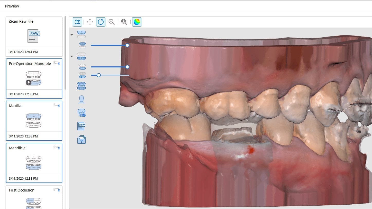
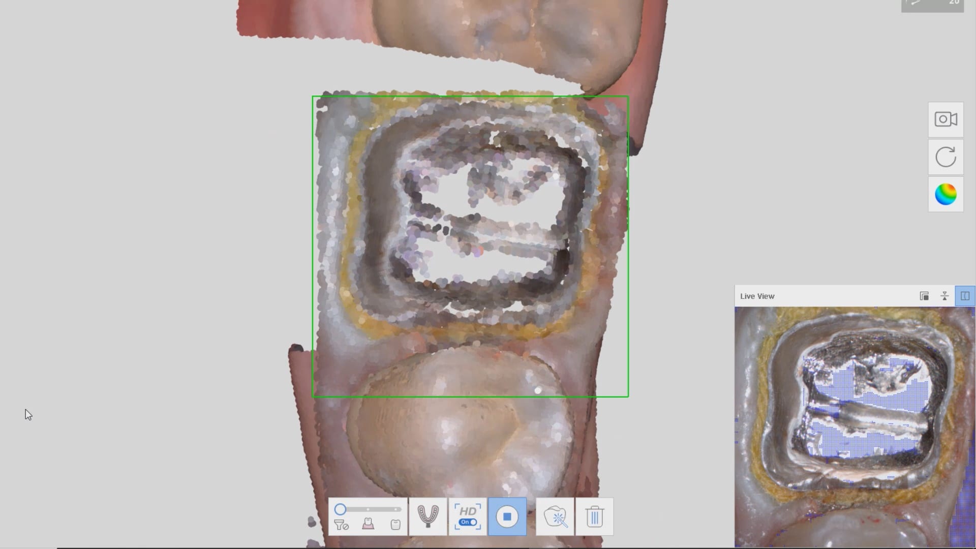
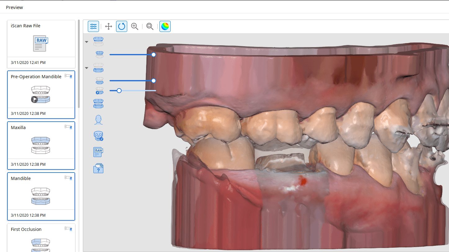
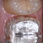
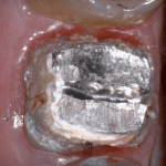
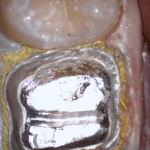
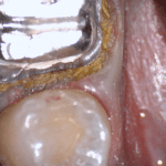
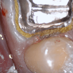
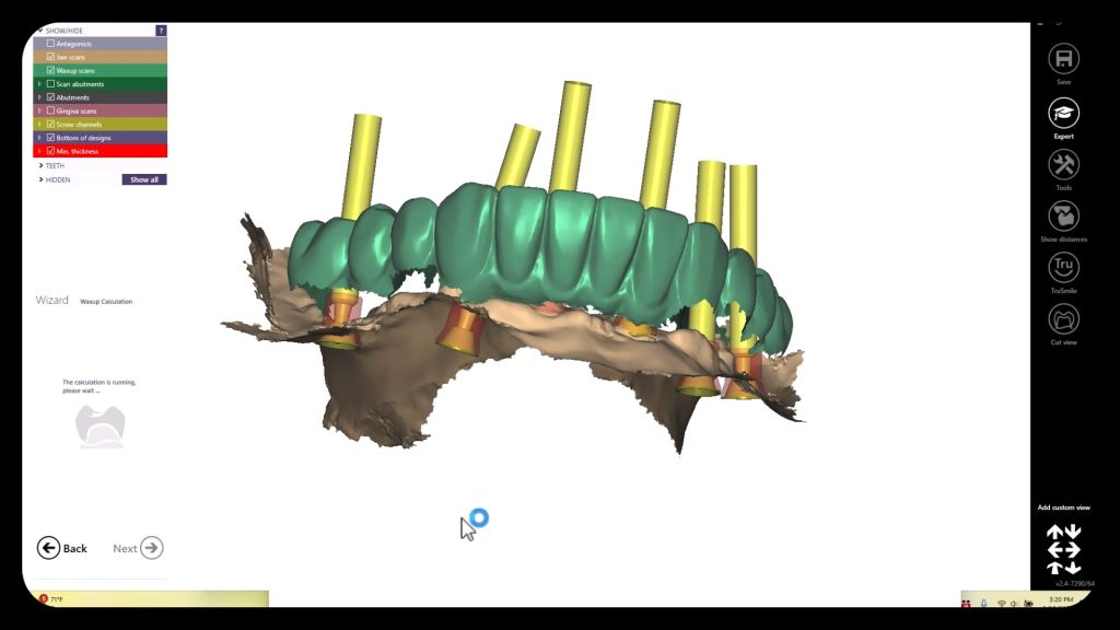
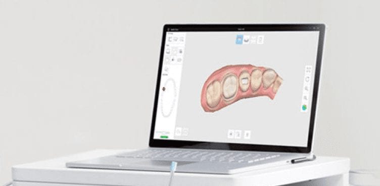
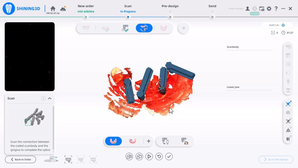
In this clinical video we demonstrate how to scan a molar preparation for the replacement of a crown with recurrent decay and open margins. The molar was root canal treated and the tissue was inflamed. the preparation was imaged and a temporary was fabricated to allow the tissue to heal properly.
The main point of this video is to show how to capture the contacts of the adjacent teeth and the deep marings




You must log in to post a comment.