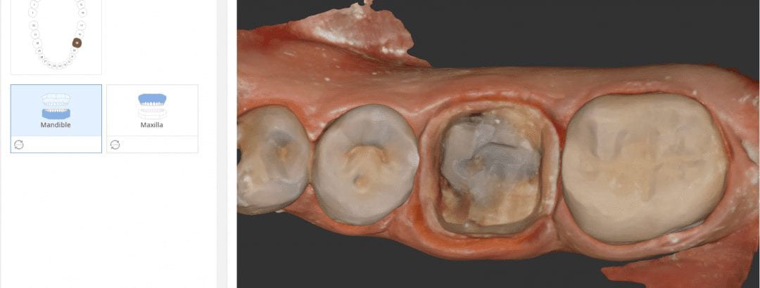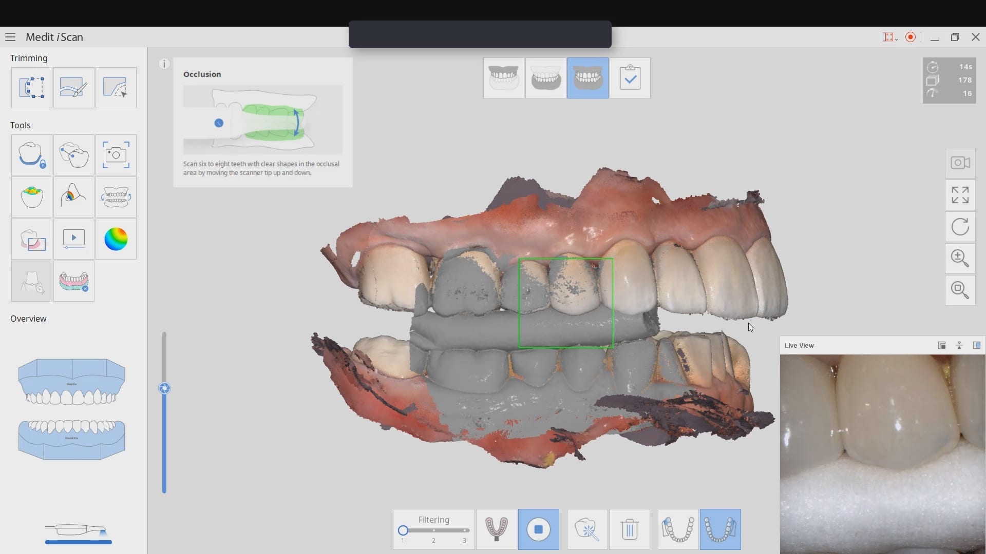
Here’s a straight forward scan of a molar crown prep. You can see how angling the camera helps you capture the contact areas of adjacent teeth. You can spend a little more time capturing more data below the height of contour, but that is not necessary as you won’t be making contact to that area (otherwise you won’t be able to seat the crown).
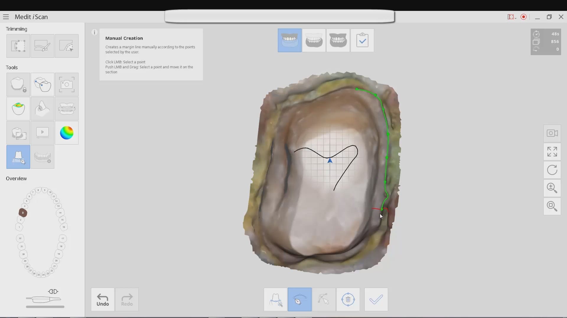
You can see the rest of the process of imaging the opposing and capturing the buccal bite and articulating the models together in the video. With the color capture, more often than not, it is not necessary to retract the tissue to visualize the margins.
[videopress ntWOMxfk]


You must log in to post a comment.