
- This event has passed.
Scanning Fundamentals and Intro to Full Digital Workflow-Las Vegas
January 17 @ 9:00 am - January 18 @ 3:00 pm PST
$400.00 – $2,500.00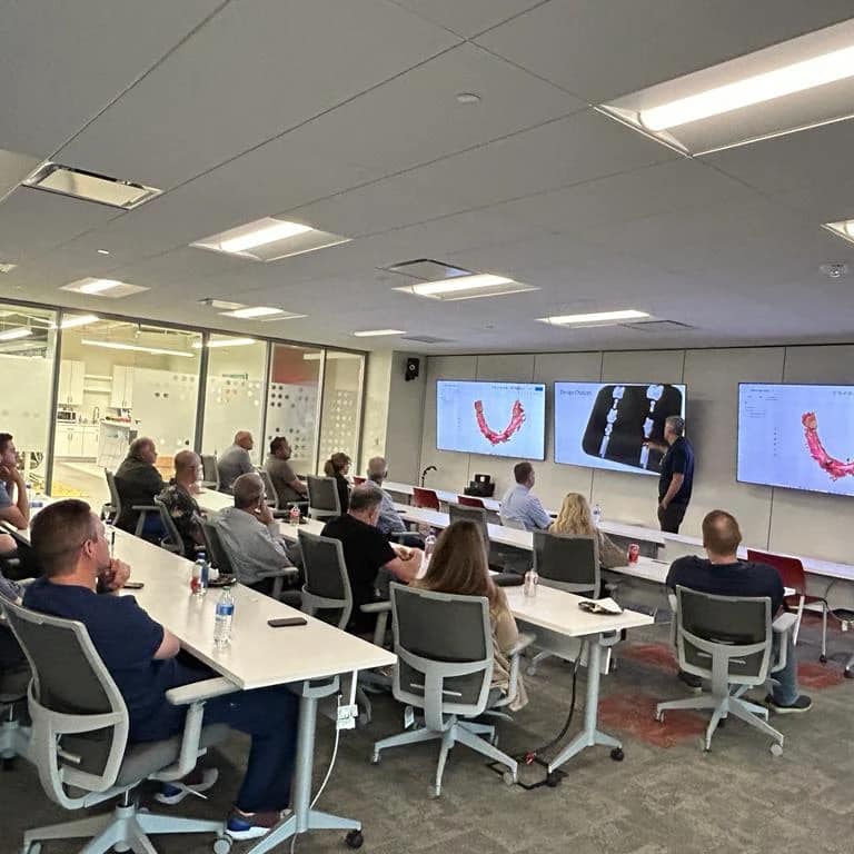
Title-Level 2-Scanning and Intro to Digital Workflow
Objectives:
- To understand the additional functionality of the digital scanner beyond simple scanning and provide insight into the procedures that can be incorporated into a practice by diving into the expanded functionality.
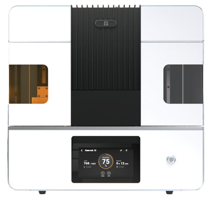
- To understand the basics of moving to an expanded or full digital workflow with the incorporation of in-house design and/or 3DPrinting or milling.
Teaching Methods:
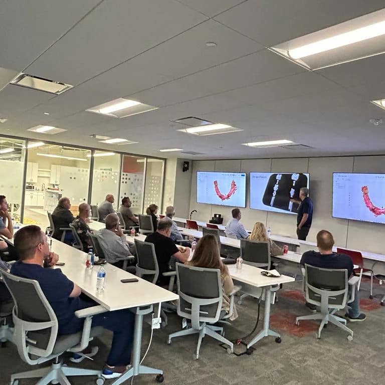
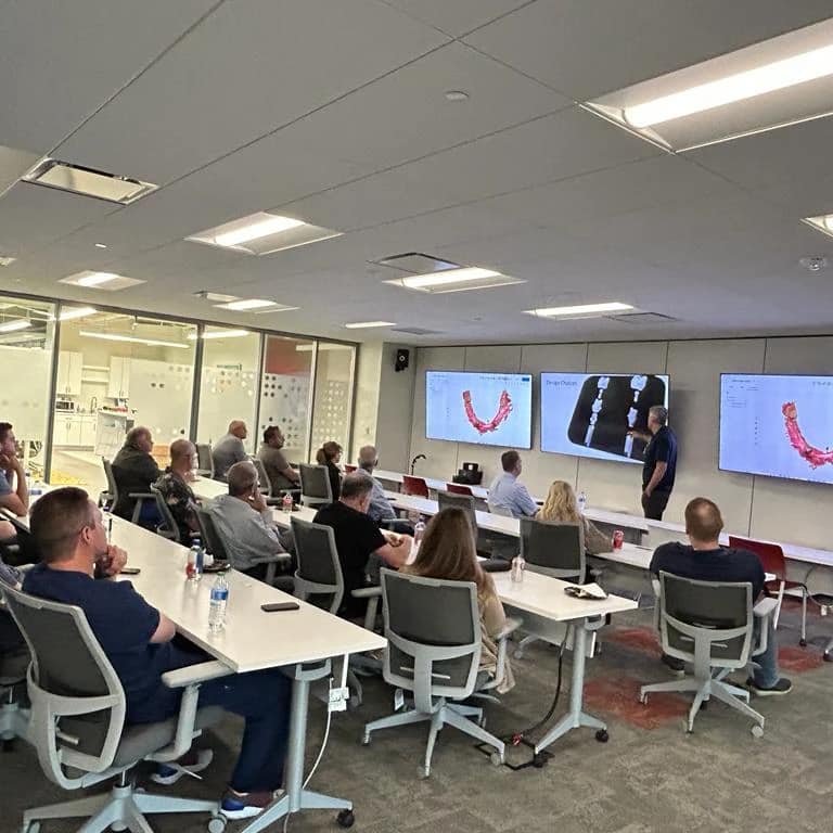
Lecture instruction, guided self-scanning and design applications, and guided computer instruction to send models to printer
DAY 1 Morning Session:
- Fundamentals of digital impressions
- Learning the advantages of digital impressions over analog procedures, functioning independently of time and sequence and taking advantage of editable and additive features of intra-oral scans
- Master single-unit impressions, complex cases and introduction to staging full arch cases
- Managing errors that arise in imaging
- Understand how to quickly capture and accurately render full arch cases
- How and what to delegate to team members
Full arch scans for aligners and other appliance can be tedious and tricky with many intra-oral scanners. With sound principles and easy tutorials, you can image a full arch in very little time. The reliability map, unique to each scanner, helps you stay on track and avoid both model distortion and inaccuracies.
Afternoon Session:
- Digital implant restorations
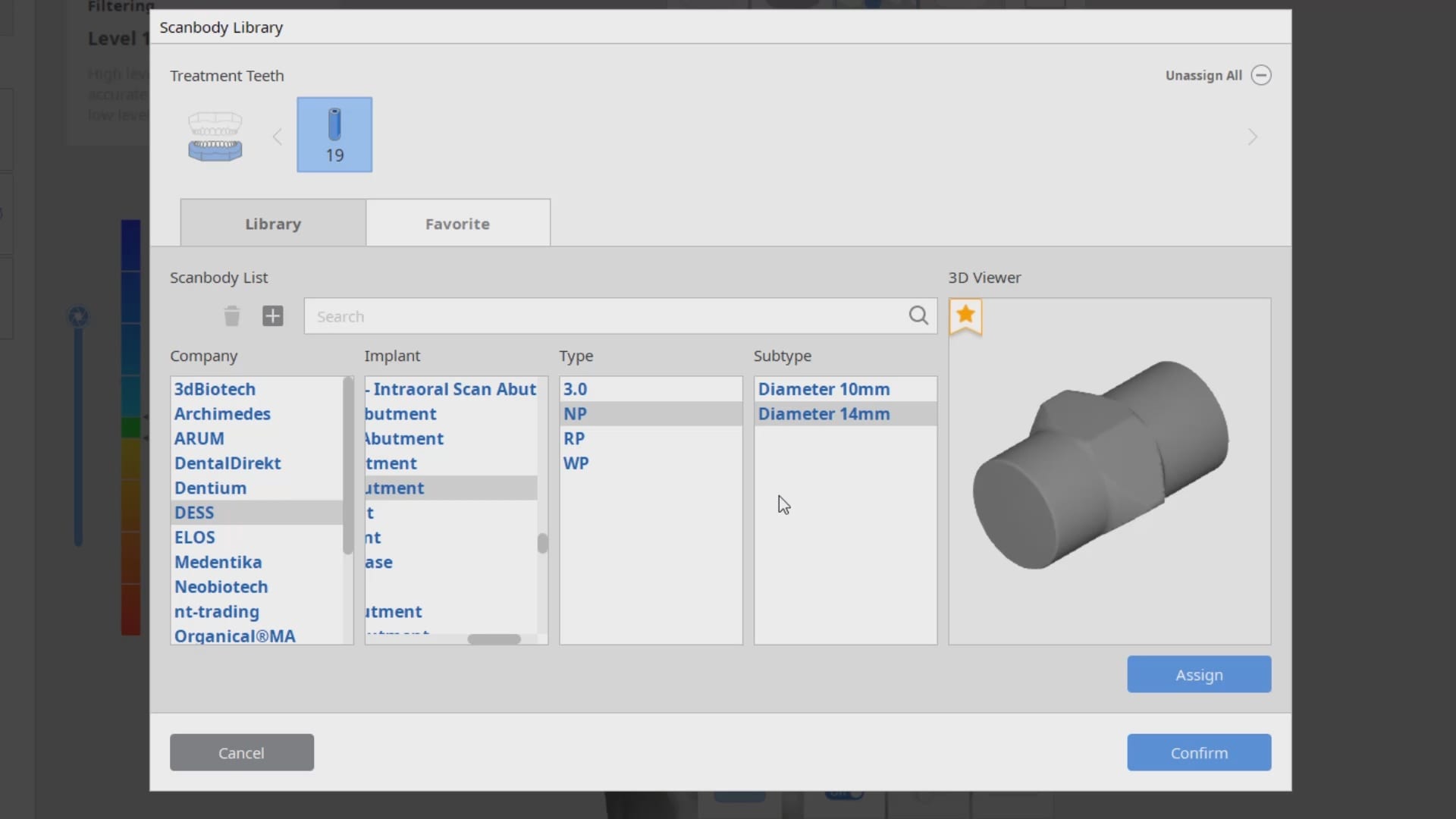
- Master the artificial intelligence implant suprastructure identification system to manage direct abutments and scanbodies
- Design single units, copy cases, and custom abutments from scanbodies
- Individual brand breakouts for a deep dive into menu functionality
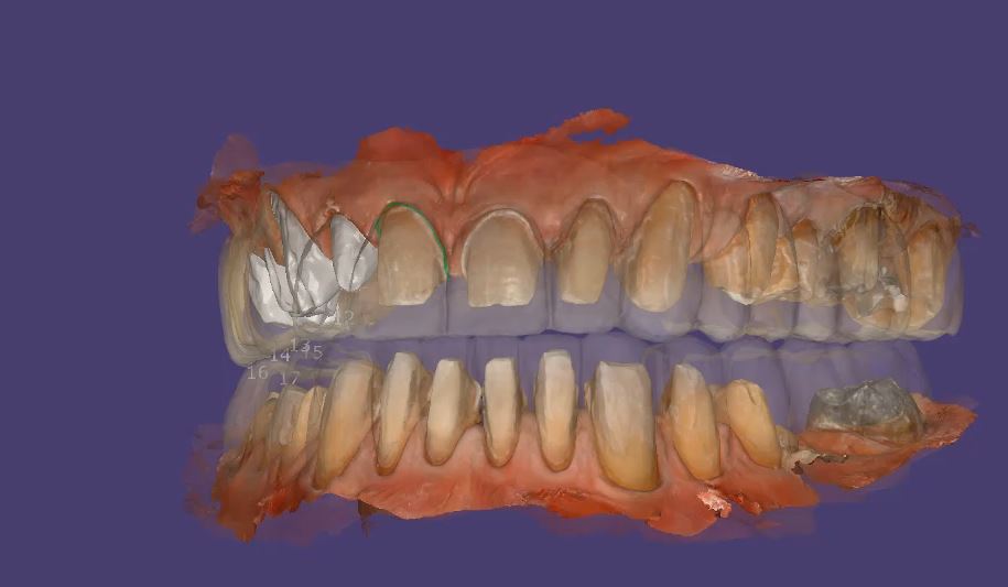
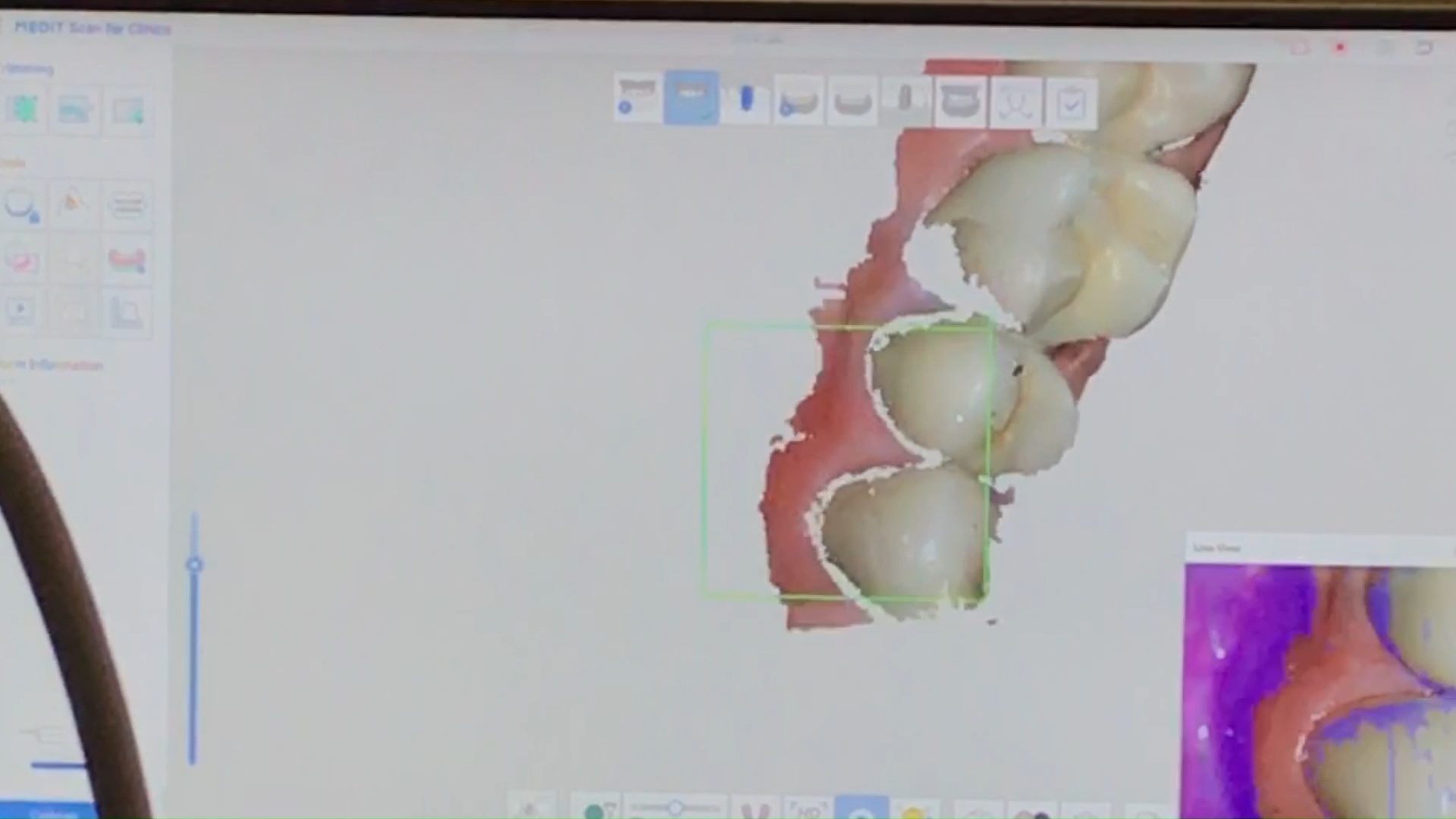
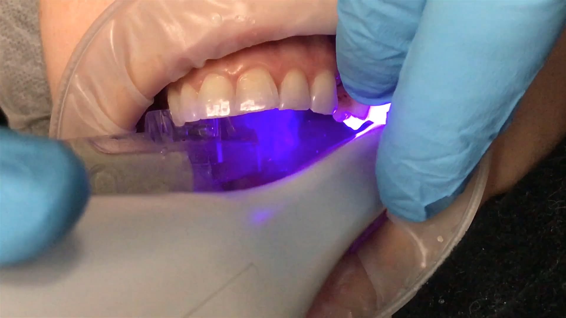
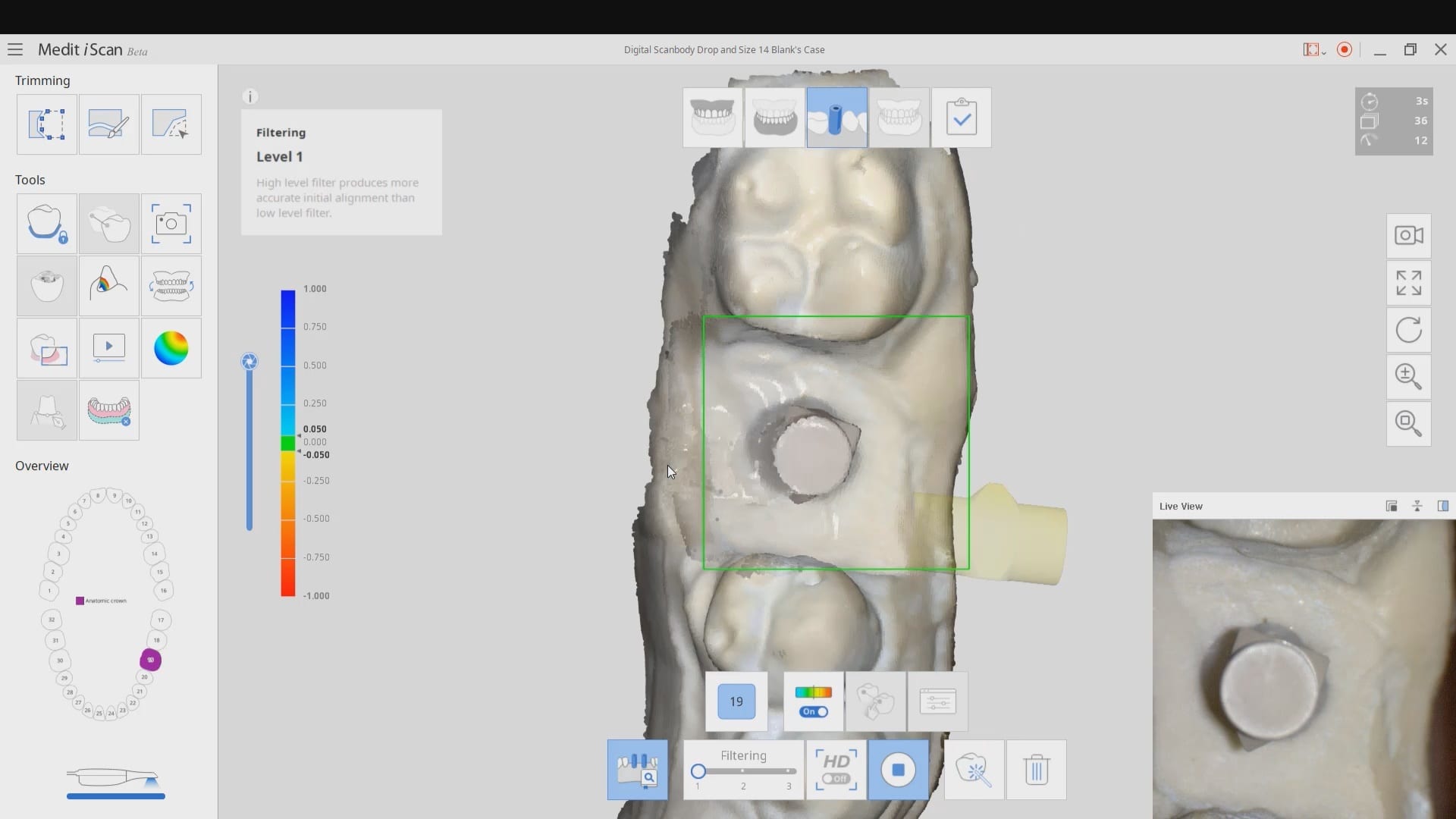
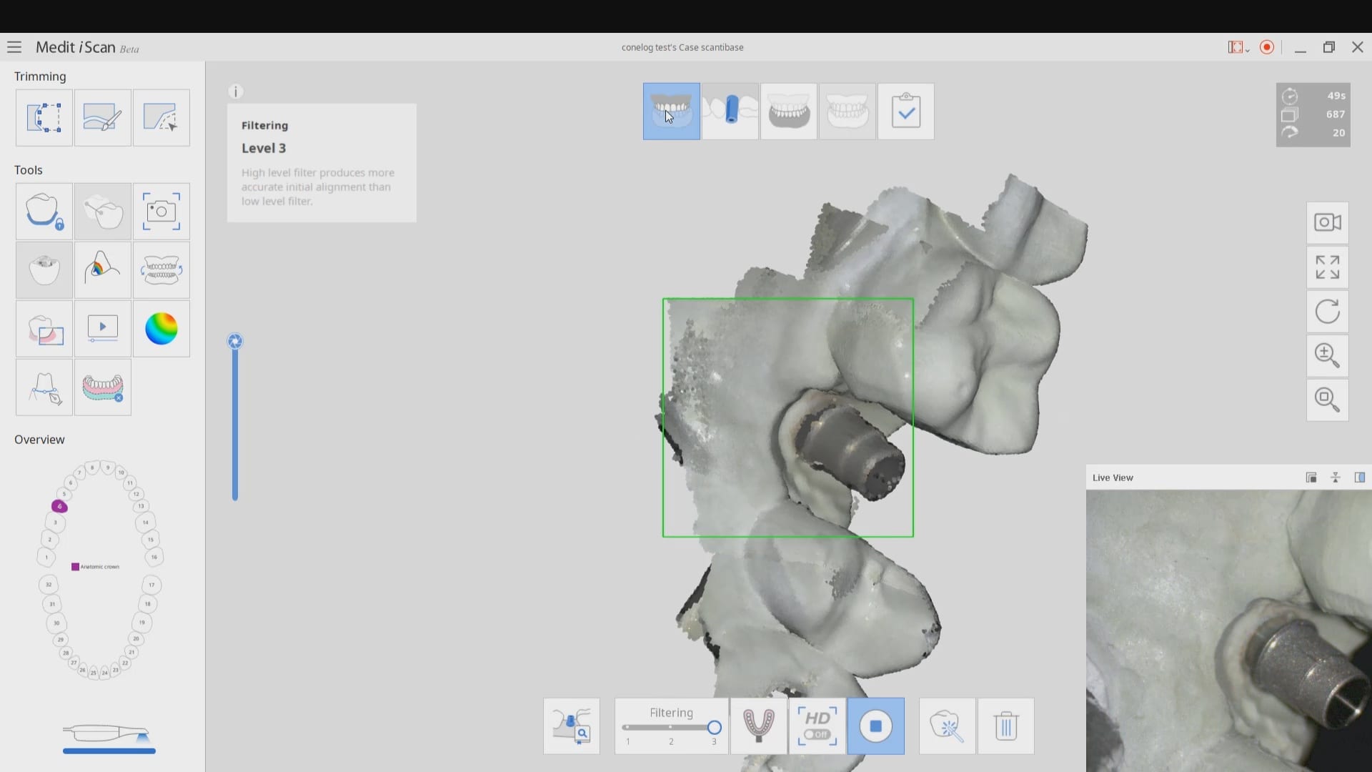
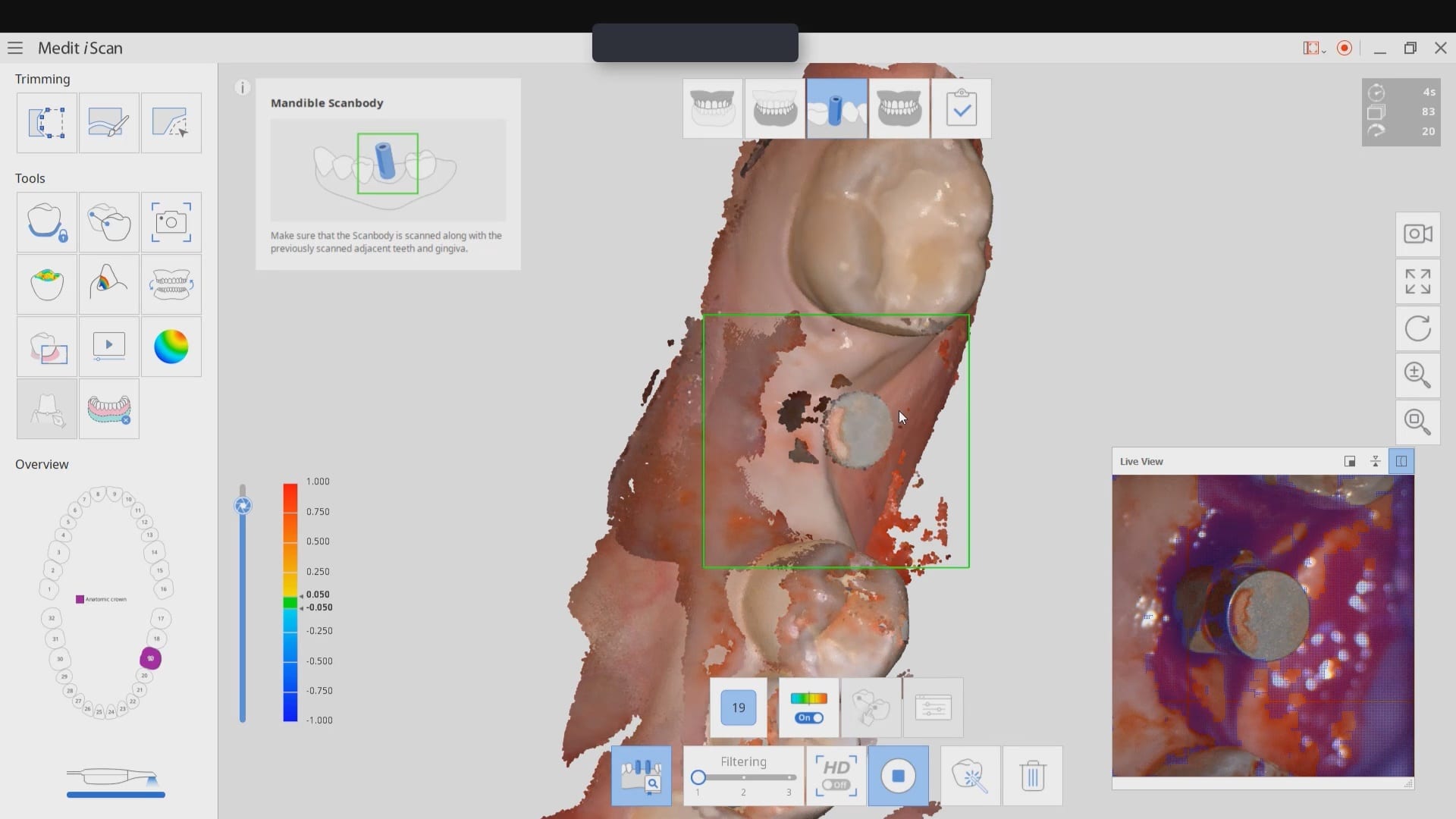
The following videos demonstrate how the scanner uses artificial intelligence to readily identify a dental implant scanbody, a direct abutment, or even an impression abutment. Even more impressive, you can import your own library of implant components or design and create your own scanbody. This automatic identification of implant suprasctructures saves dozens of steps, whether you wish to fabricate restorations in the office or send it to the lab.
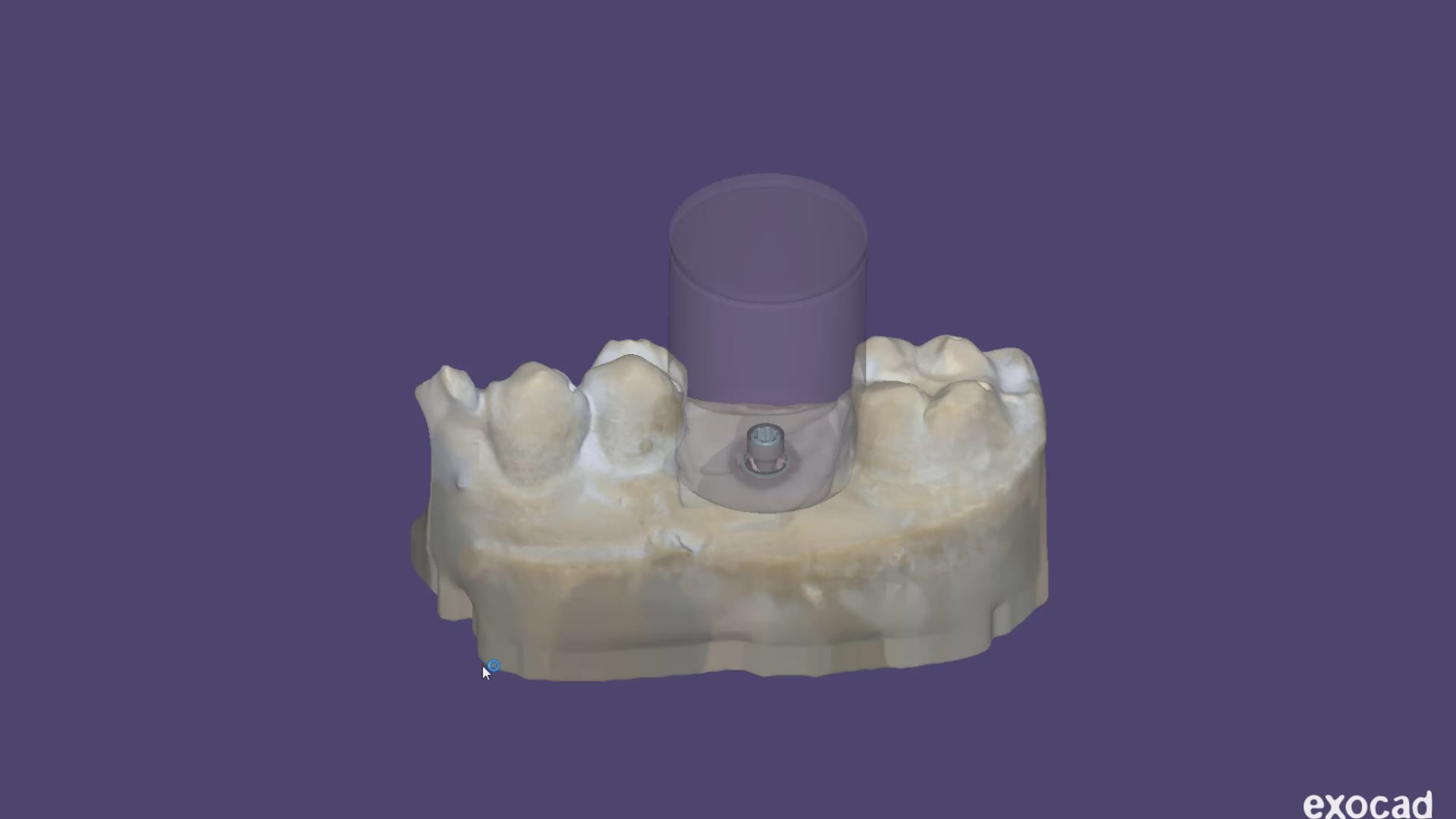
The scanner’s artificial intelligence implant identification system spots a titanium scanbody on a freshly uncovered biomax implant. It didn’t matter how bloody the field was. The scanner readily identified the suprastructure, which is then transferred to the cad software (where the implant location and timing is correctly displayed) and the design of the custom abutment begins. This process is much more efficient and reduces potential errors. Another amazing feature is that the software can recognize impression abutment, tibases, and lots of other supra structures.You can see this case in more detail here
DAY 2 Morning Session: Digital Implantology
- Introduction and mastery of Integration of Cone Beam data and STL data
- Understanding the limitation of Cone Beam and STL integration
- Integration and limitation of multiple Cone Beam data sets
- Proper digitization of denture duplicates for stent fabrication
- Multiple ways to design a surgical stent and how to choose appropriate design type
- Proper case presentation techniques for patients
- Single implant placement designs
- Understand how to use Cone Beam scans and surgical stent
- Treatment planning large cases
- Appropriate case set ups and their ramifications during surgery
- The multiple uses for chairside denture duplication
- Chairside denture duplication and scan appliance fabrication
- Impression techniques for multiple fixtures
- Impression techniques for multi-abutments
- Restorative designs of abutments on CAD software
Afternoon Session: Edentulous and Full Mouth Cases
- Immediate extractions and how to keep track of the vertical dimension
- The multiple restorative options available to you
- Accurate transfers of fixtures to models
- Digital impressions for edentulous cases
- Edentulous case design with digital dentures
- Photogrammetry and intra-oral scans of full arch cases
CE Credits: 14
Presenter-Dr. Armen Mirzayan
Armen Mirzayan graduated from Northwestern Dental School in 1998 and completed his General Practice residency at the Queen’s Medical Center in Honolulu, Hawaii. He met his wife, Dr. Jean Lee-Mirzayan in dental school and they started a practice together in downtown Los Angeles.
Early in his career, Dr. Mirzayan believed that there was little chance that he (and the industry as a whole) would continue to practice analog dentistry for too long, and he eventually purchased a CEREC system in January of 2001. Since then, he has used every version of the CEREC, and has milled on more than a dozen different chairside milling machines. He is also well versed in CBCT technologies and has had a unit in his own practice since 2009. Since then, he has trained well over 10,000 dentists in the use of CAD/CAM, CBCT and guided implant technologies.
In addition to private practice, he co-founded www.cerecdoctors.com; the original site that fostered a training/social network for the community of digital dentists, and essentially set the tone for the growth that the industry enjoys today. In 2013, Dr. Mirzayan founded CAD-Ray to serve as a center for teaching dentists guided surgery techniques and how to fabricate surgical guides. Since its inception, CAD-Ray has manufactured over 35,000 surgical guides and counting. It wasn’t until 2018 that the distribution arm of CAD-Ray was formed, which focuses on the distribution of reliable, efficient digital dentistry options as well as focused training and support for those technologies.
These days, Dr. Mirzayan spends most of his professional time in three ways. First, he travels the country teaching intraoral scanning, digital dentistry shortcuts and advanced dental techniques in his role as the clinical education director for CAD-Ray. Second, he rigorously tests new digital (dental) equipment; evaluating each product for reliability and durability to see if they are worthy of joining the CAD-Ray portfolio of supported products. Third, he still practices dentistry; both as a way of staying fresh, and also to test out new techniques and product innovations. His favorite part of the business is knowing that the strong majority of practicing CAD-Ray customers use their equipment on a daily basis.
Related Events
We offer a variety of courses focused on Digital Dentistry:
Level 1 – One Day Regional Intra-Oral Scanning by one of our mentors. Introductory level courses that give you and a team member the confidence to scan an arch in under a minute.
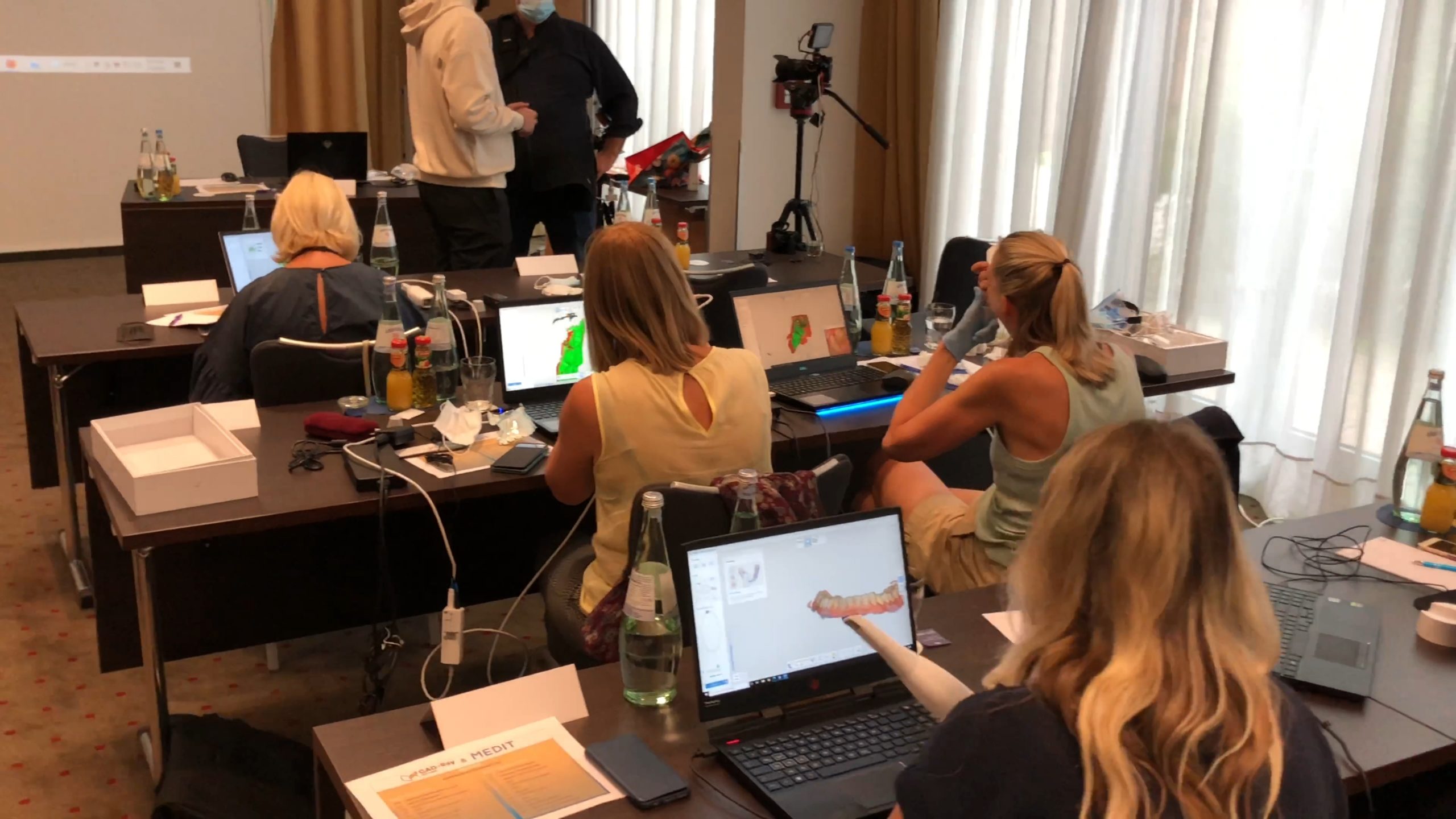
Level 2– Two day hands-on training course for Medit i500 users on digital imaging and unleashing its full potential
Level 3 -Two day hands-on training on digital restoration designs and manufacturing, utilizing our CAD-Ray Design Software and all of your milling options, including milling titanium abutments or ceramics to tibases
Level 4 – Two day hands-on Digital Implantology Course from guided surgery to iCam and Medit Training and on implementing CT technology into your practice, focusing on guided surgery and apnea treatment
We frequently attend conferences and trade shows around the country
Advanced Technology Center is a Nationally Approved PACE Program Provider for FAGD/MAGD credit. Approval does not imply acceptance by any regulatory authority or AGD endorsement. October 1, 2019 to September 30, 2023 Provider ID# 337070
CAD-Ray Event Cancellation Policy
CAD-Ray plans classes well in advance, so funds are usually captured for the preparation and arrangement of venues, food, materials and marketing for each event. In the event that an attendee must cancel, we ask to be notified at least 24 hours prior to the start of the event, or CAD-Ray reserves the right to keep the attendance fee to cover non-recoupable expenses already laid out by the organization.





You must log in to post a comment.