Settings Option During Imaging
Lessons
Purple Silhouette in Live View is an Indicator of Objects the Medit i500 Cannot See
The new Medit i500 software, version 2.0, gives you a variable setting for your depth of view. You can use this to your advantage when scanning certain cases, as long as you appreciate what is happening in the live view mode, in the lower right corner of your capture screen The purple shine on tooth […]
High Resolution Scanning
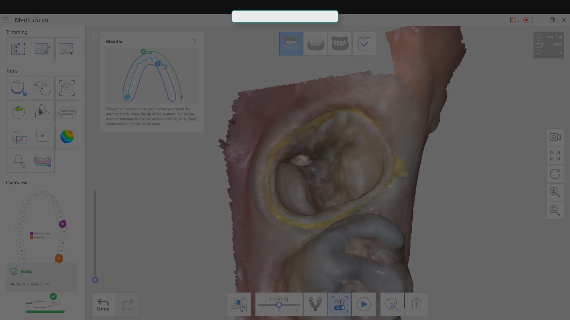
High resolution scanning allows you to pick up a lot of detail when needed. Although it dramatically reduces scanning speed, it gives you the detail you are looking for when […]
Focal Length of the Medit i500 IOS in action
With a focal length of -1.5 to 17 mm’s as of August 6, 2018, we took the scanner for a test drive. This particular clinical situation has a failing implant fixture in the site of the first molar, where there is bone loss to the third or fourth thread. The patient had a full mouth […]
12 mm focal length setting, optragate, and isolite

As a new user, you may find the optragate and the isolite of tremendous help when imaging a quadrant. The hardest area to image is the lower second molar. In this video we show you some methods where you can reduce the focal length to 12 mm to ignore the tongue and the lip while […]
Deep and Bloody Scans with the Medit i500 Blue and White Cameras
The Medit i500 V2.0 has two new features we are beta testing for Deep Scans and for red / bloody environments. There is a toggle bar that allows you to […]
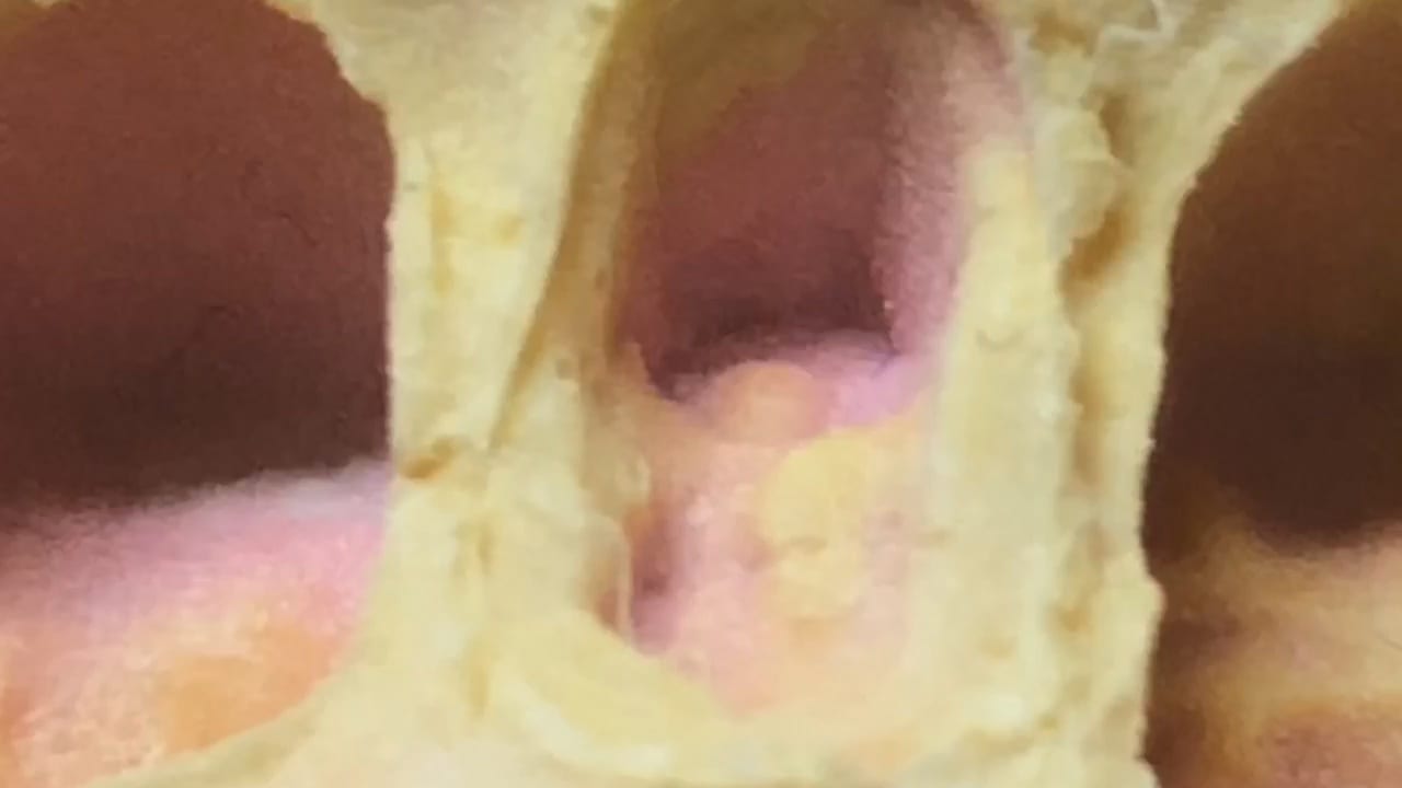
Imaging a Triple Tray Impression with Medit i500
Imaging in Model Display Mode
An advanced method of imaging is to use the model display mode. Most users focus on capturing all the data and forming a 3D model. This mode, lets you know […]



You must be logged in to post a comment.