Posted on Leave a comment

Proving the Accuracy of Shining Elite Intra-Oral Photogrammetry Scanner

Here is a quick introductory video of how the process works and the subsequent cases provide a lot more detail and information

video
Quick Overview

play-rounded-fill play-rounded-outline play-sharp-fill play-sharp-outline
pause-sharp-outline pause-sharp-fill pause-rounded-outline pause-rounded-fill
00:00
01:42

Case 1

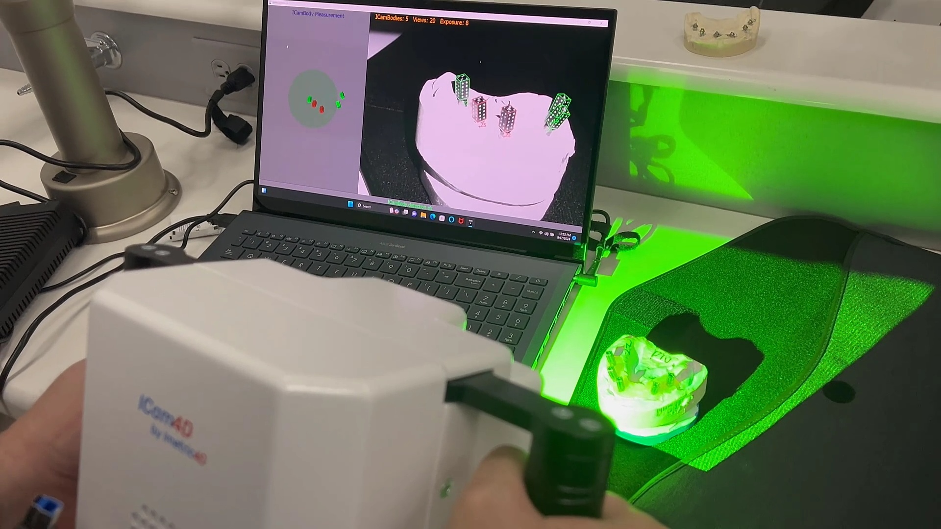
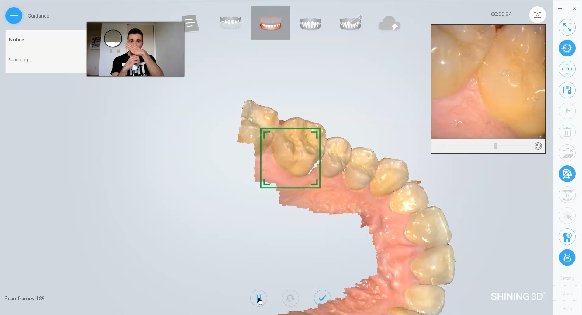
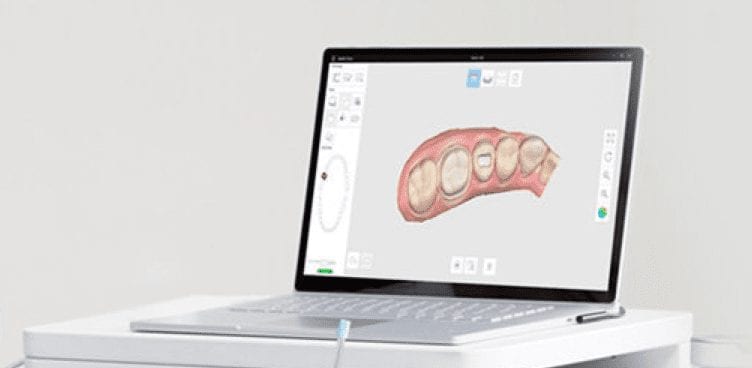
In this video we detail how the process works and how you capture everything you need for your full arches with one scanner.

video
Details of Case in the Overview

play-rounded-fill play-rounded-outline play-sharp-fill play-sharp-outline
pause-sharp-outline pause-sharp-fill pause-rounded-outline pause-rounded-fill
00:00
06:47
Item 1.Item 1.


Precision, Trueness, and Accuracy

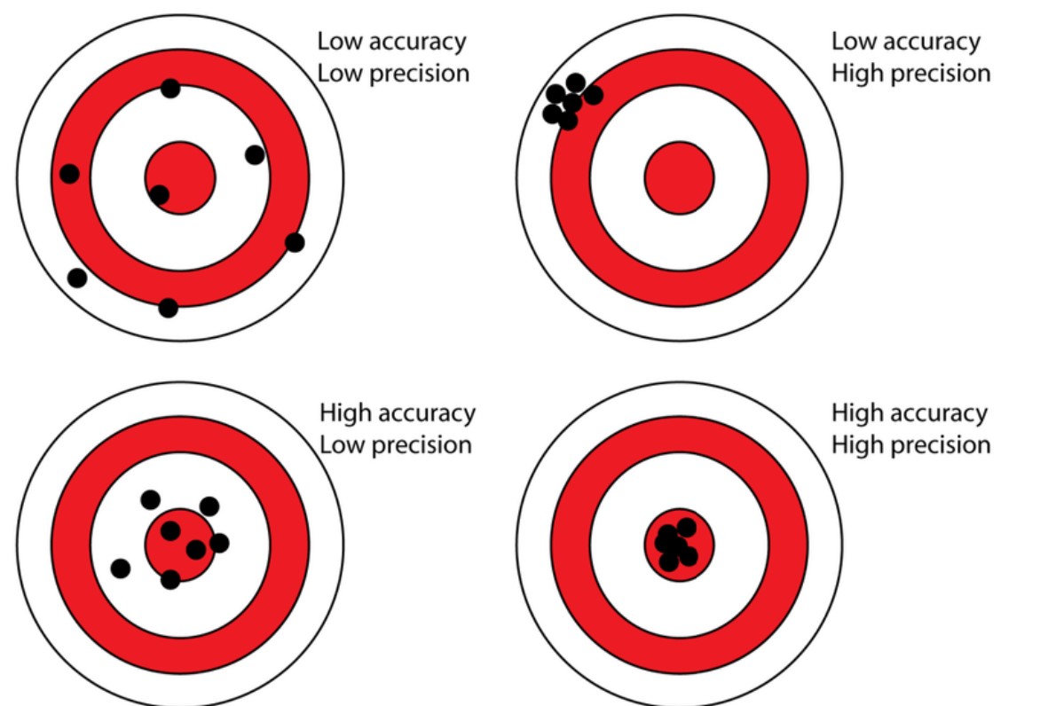
A common property that is oftentimes used to describe how well scanners perform is how accurate, precise, or true it is.  A lot of people use this incorrectly to describe intraoral scanners and it is utilized in poor judgment because there are too many variables in the input of the equation to ascertain any conclusion from the output file.  But these characteristics CAN be used to study photogrammtery.

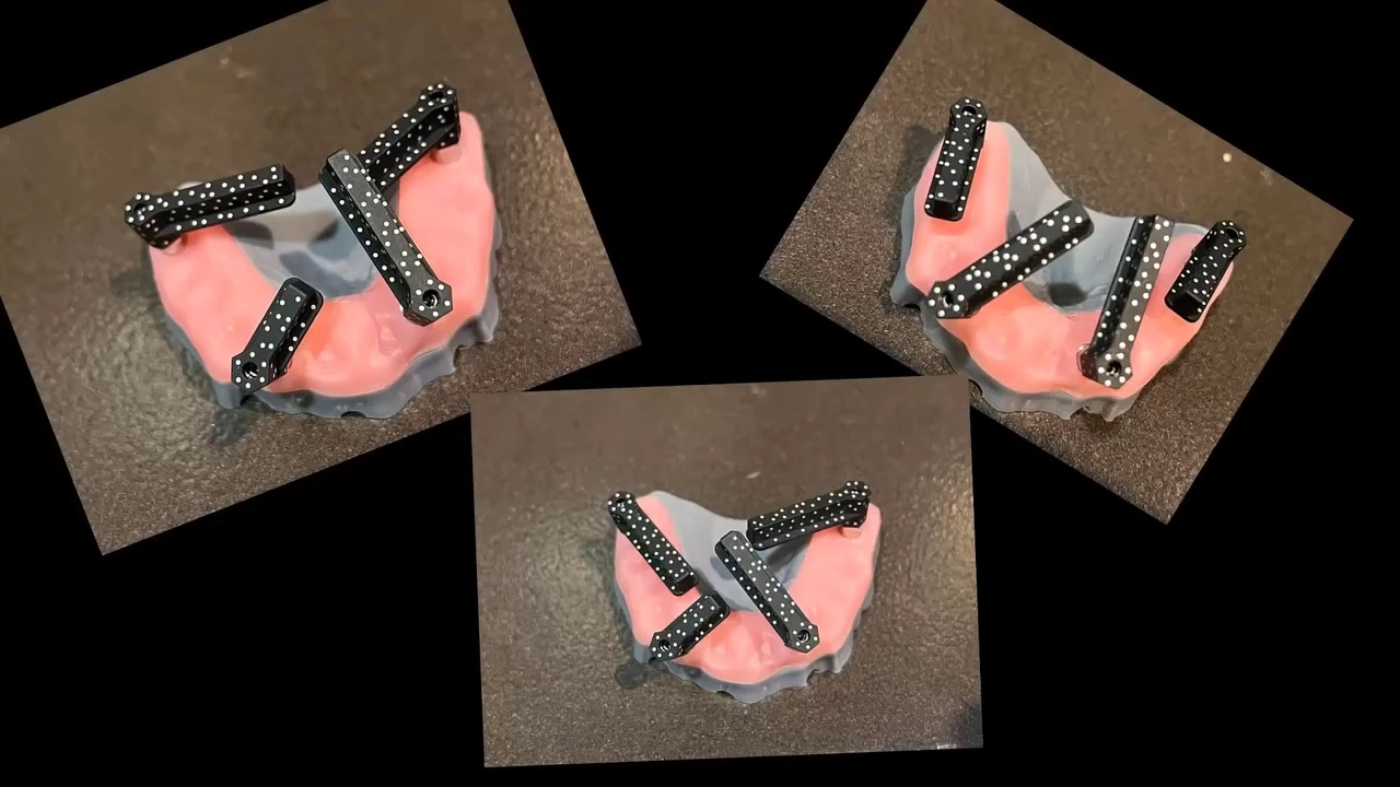
In this chart, the bullseye represents the true value (true location of the object), while black dots represent measurements (the estimated 3D locations of the object based on the 2D images) from this source.   To measure the precision, we took 3 random arrangements of the dominos and found the output files to be identical.  To measure its accuracy and trueness, we related it to a desktop scan of the same model

Precision Test

play-rounded-fill play-rounded-outline play-sharp-fill play-sharp-outline
pause-sharp-outline pause-sharp-fill pause-rounded-outline pause-rounded-fill
00:00
04:08
Item 1.Item 1.

you can download these models and test them out for yourself:  compare elite to icam and desktop scan

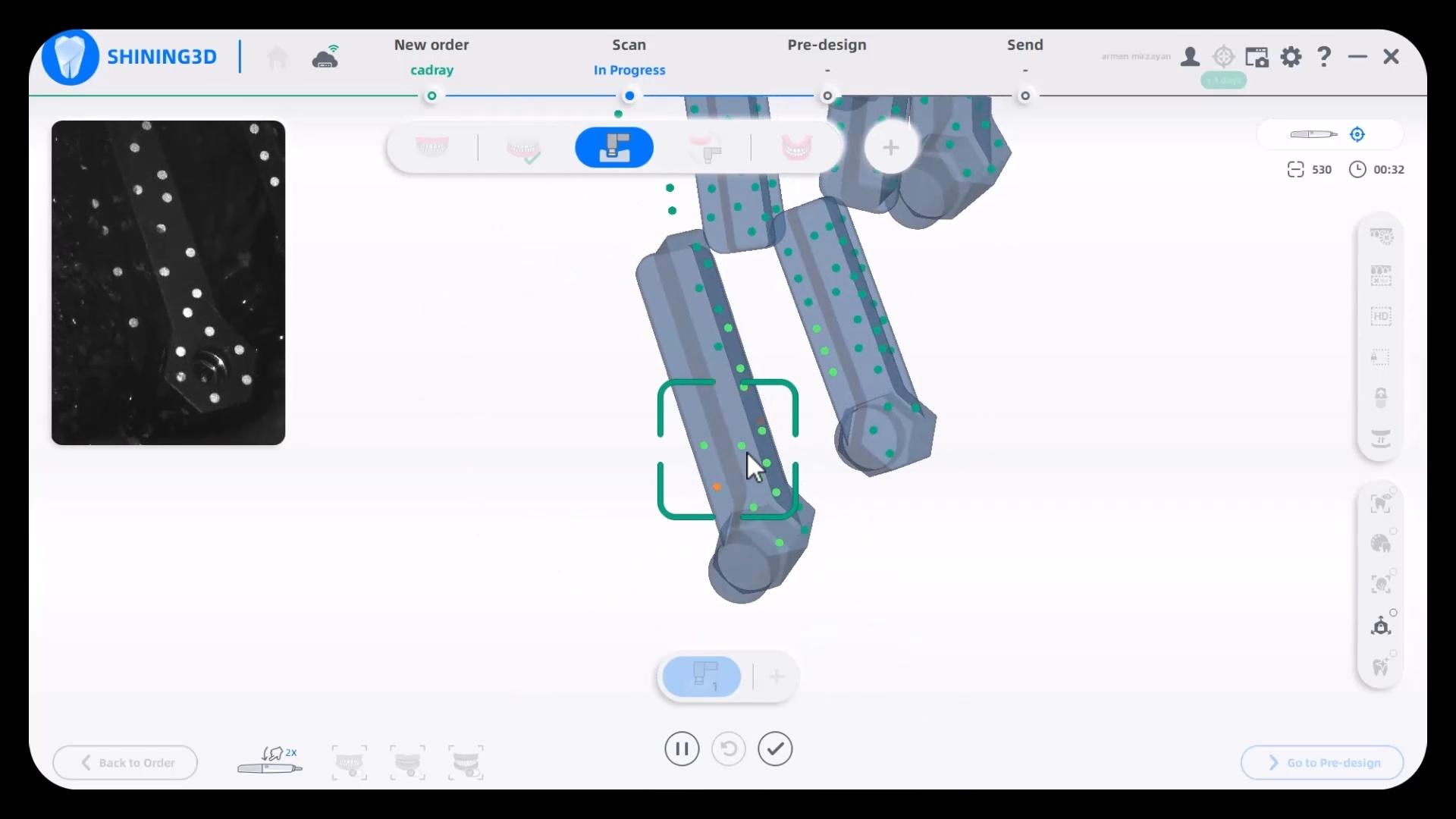
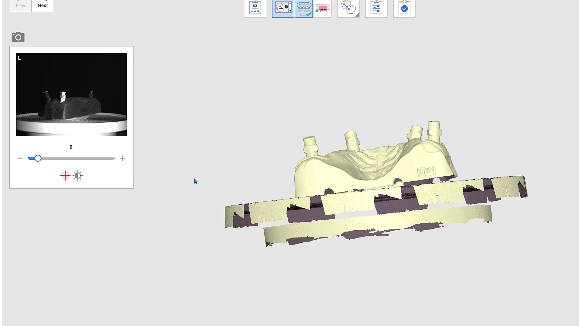
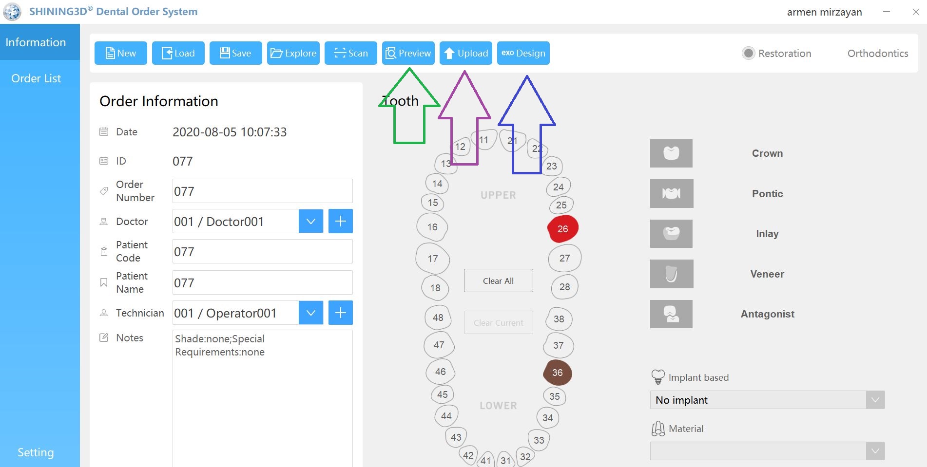
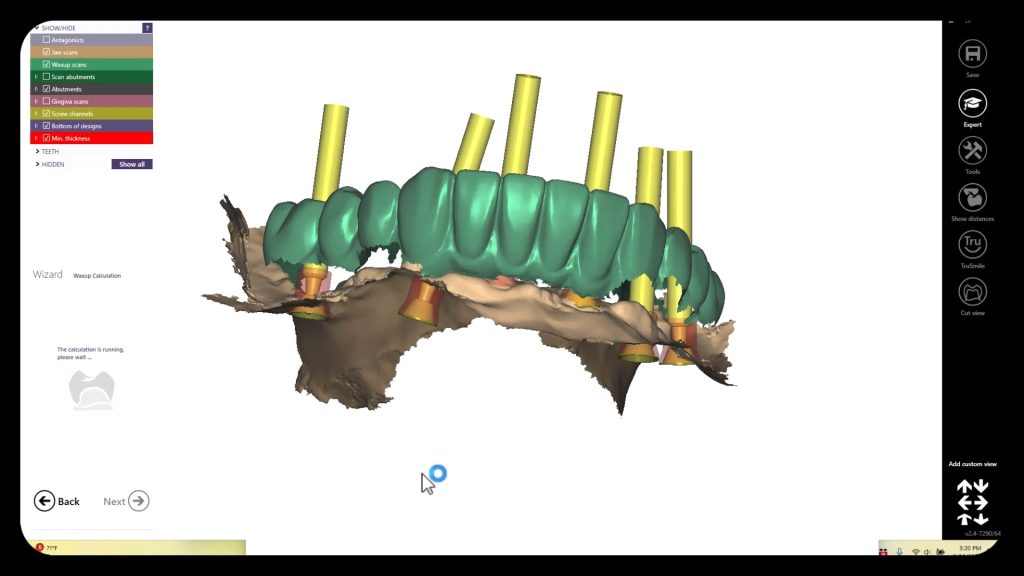
When is image capture complete?

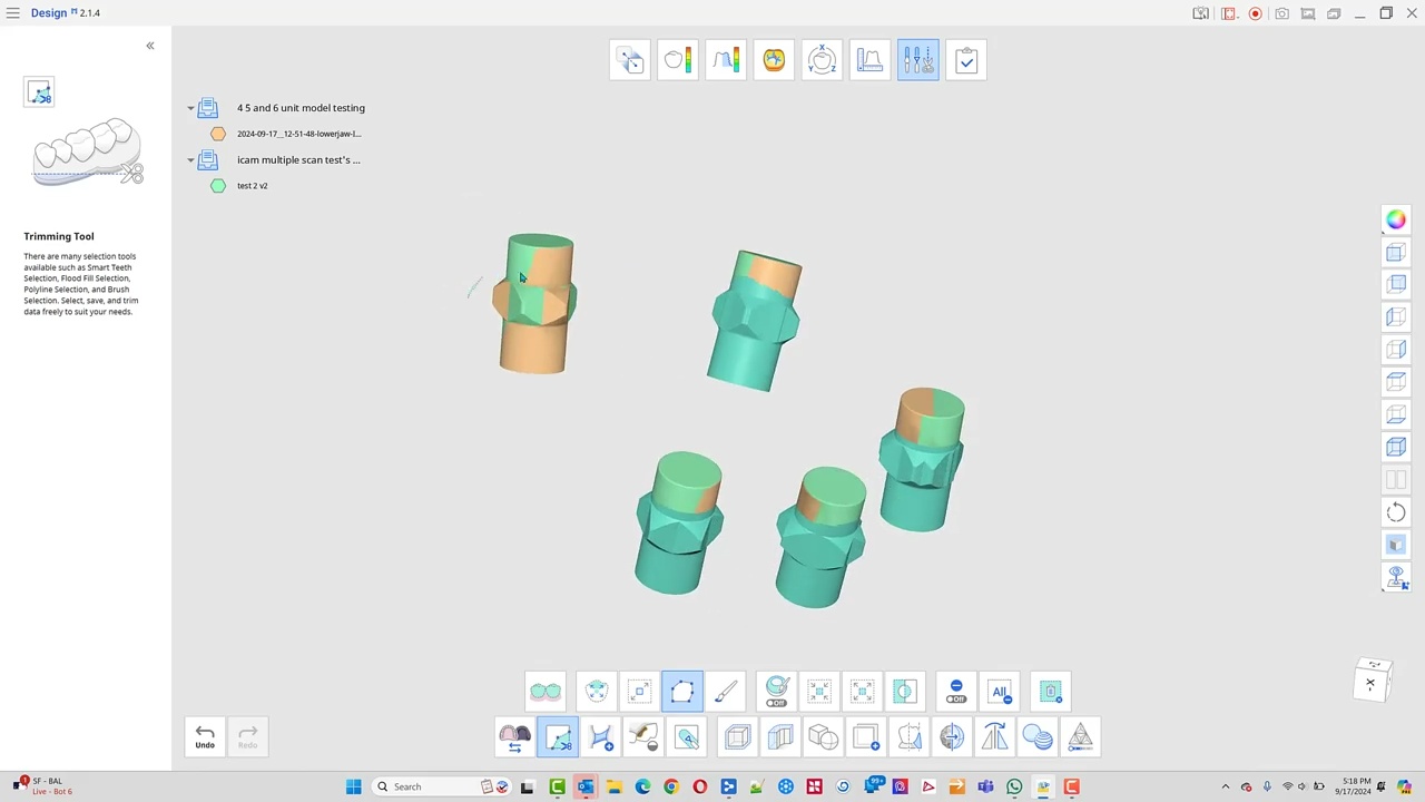
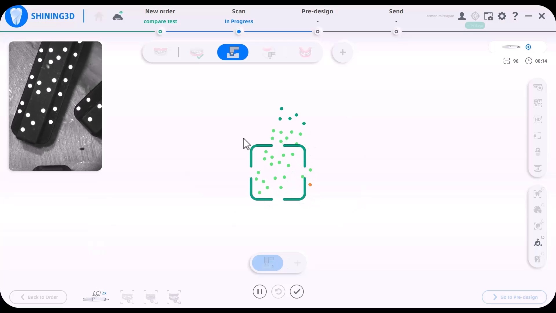
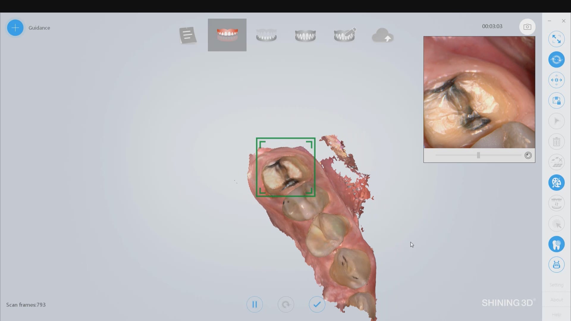
A frequent question we field is how does an operator know how much of the dominos / scanflags to capture.  The manufacturer recommends a specific scan path and to completely capture the suprastructure.  Our internal testing shows that there is no difference between the results as long as you allow the software to spit out the scanflag geometry while you are scanning. See this video for an explanation

video

play-rounded-fill play-rounded-outline play-sharp-fill play-sharp-outline
pause-sharp-outline pause-sharp-fill pause-rounded-outline pause-rounded-fill
00:00
04:44

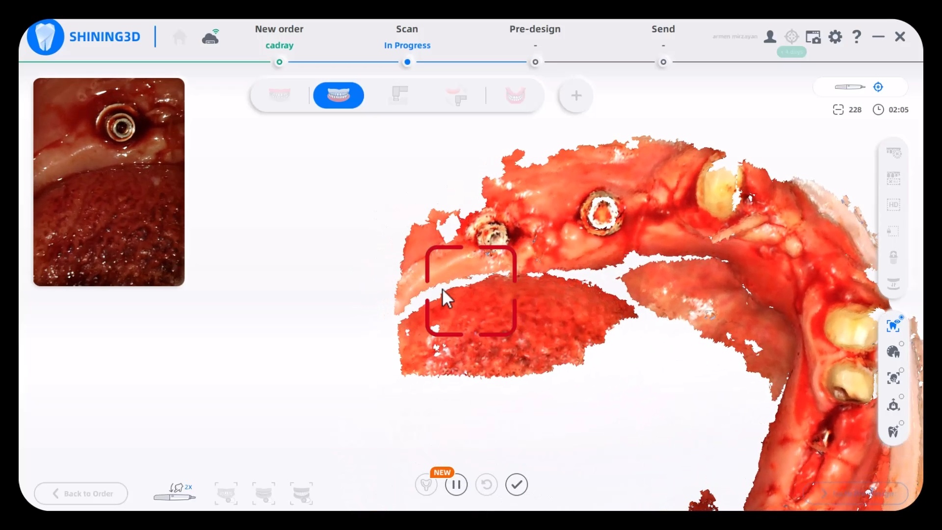
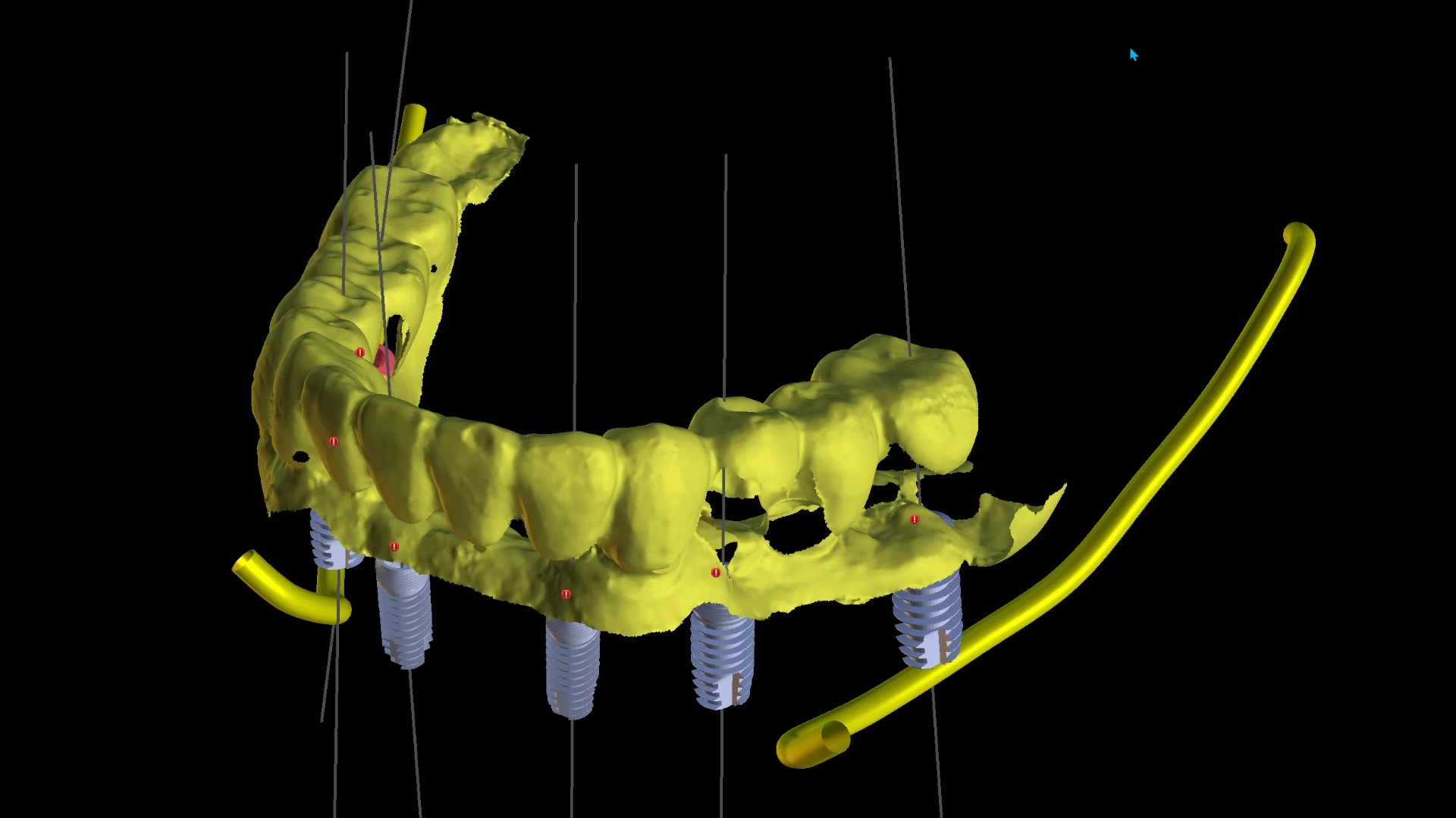
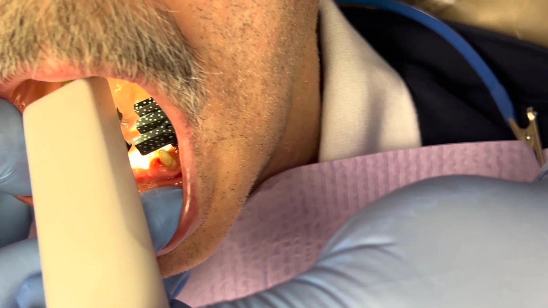
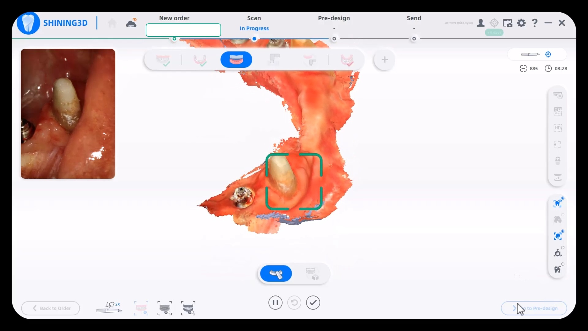
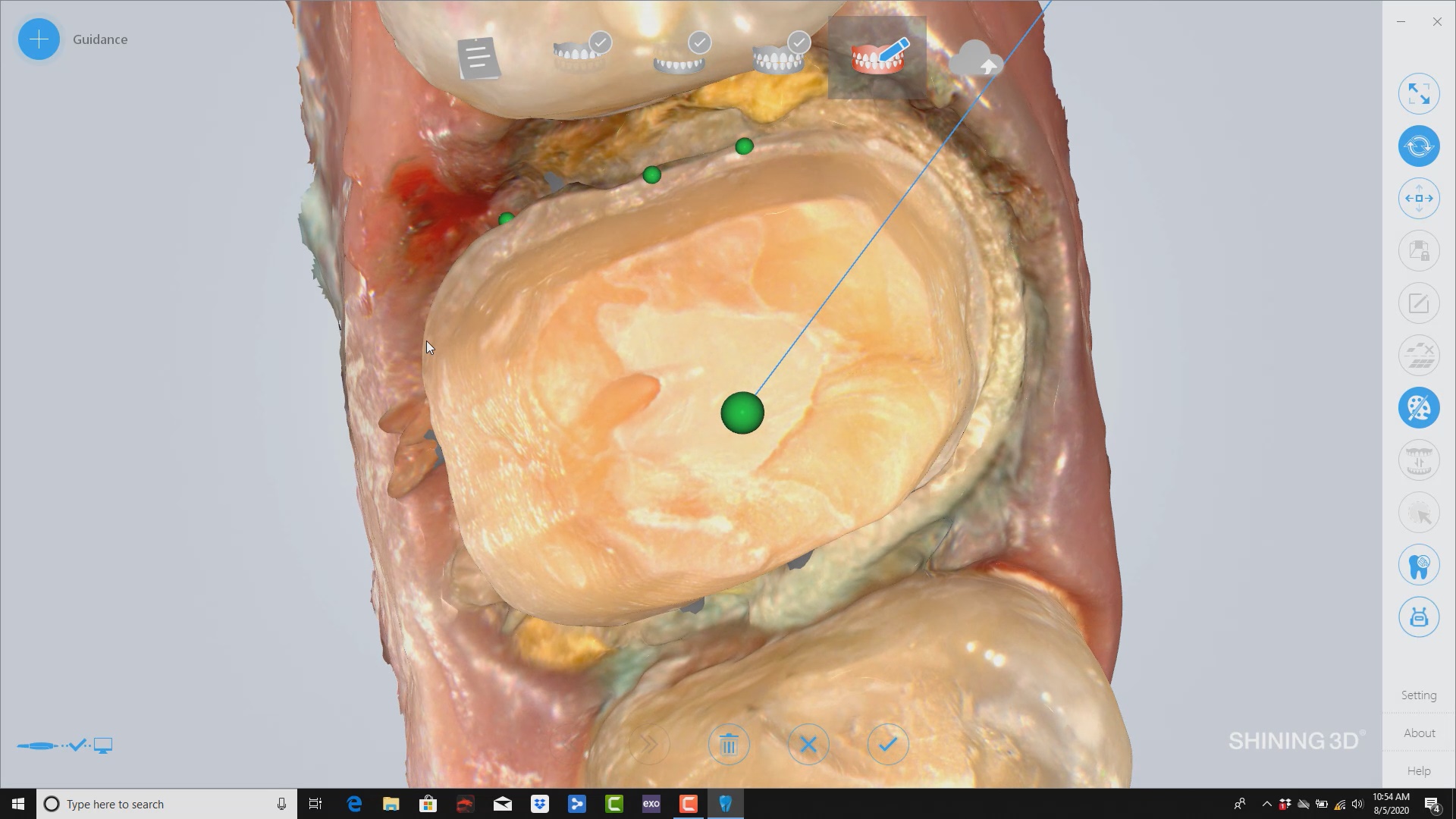
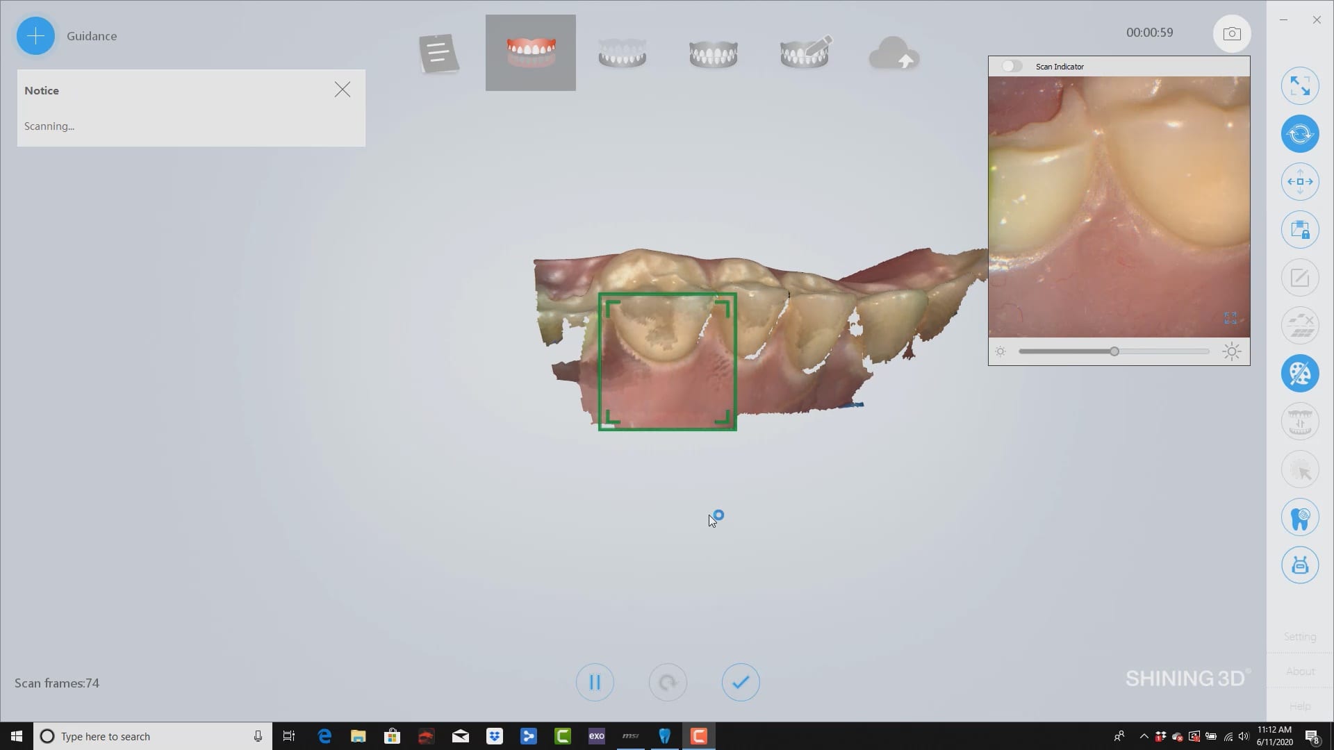
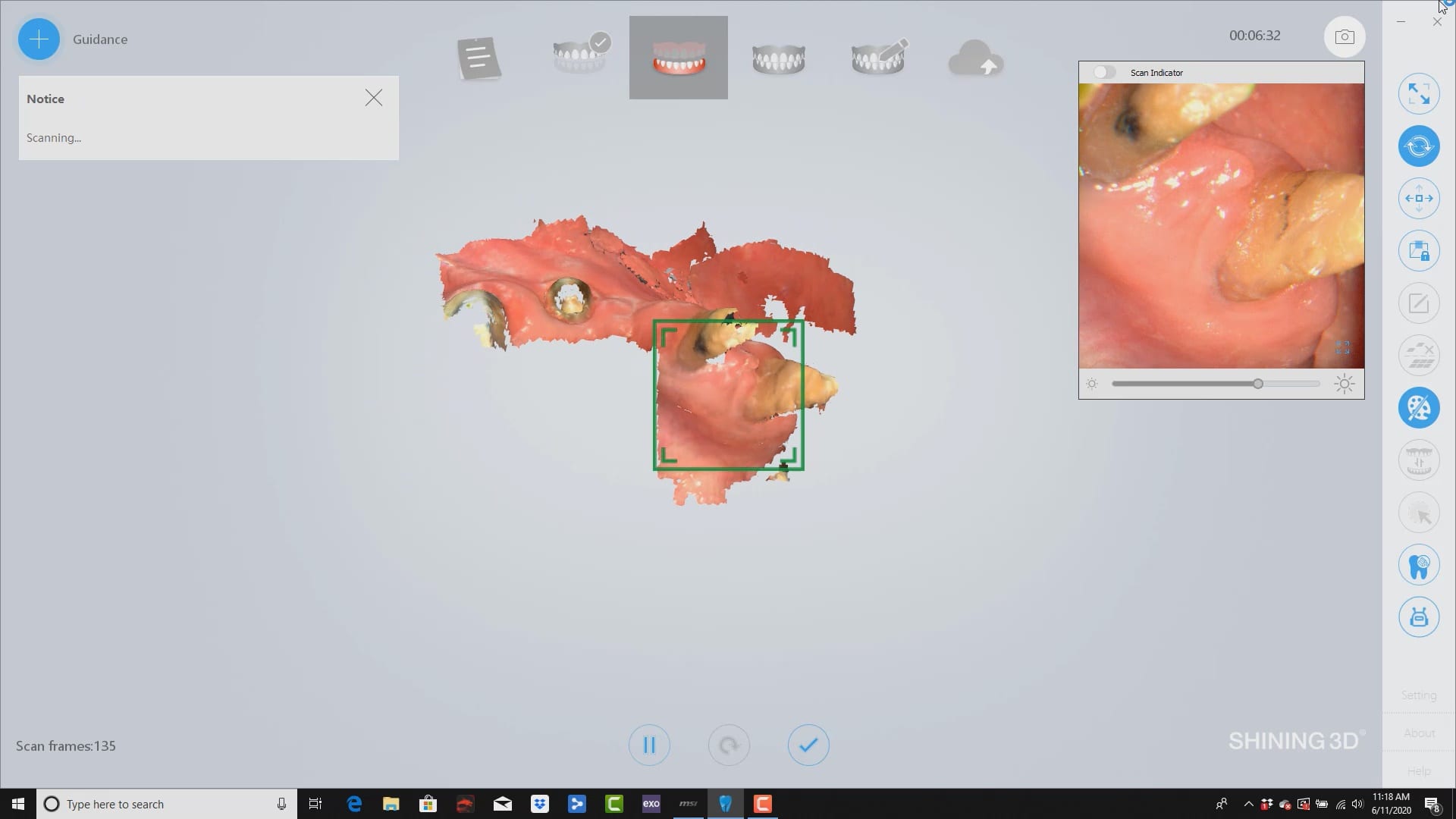
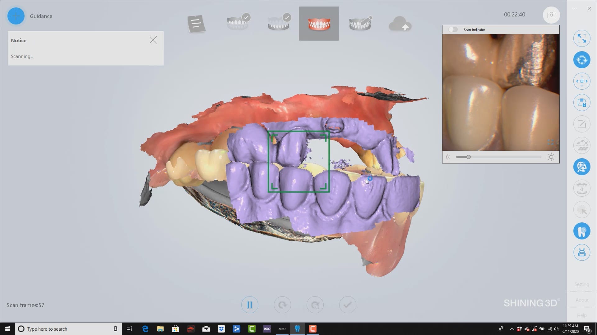
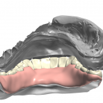
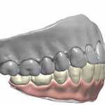
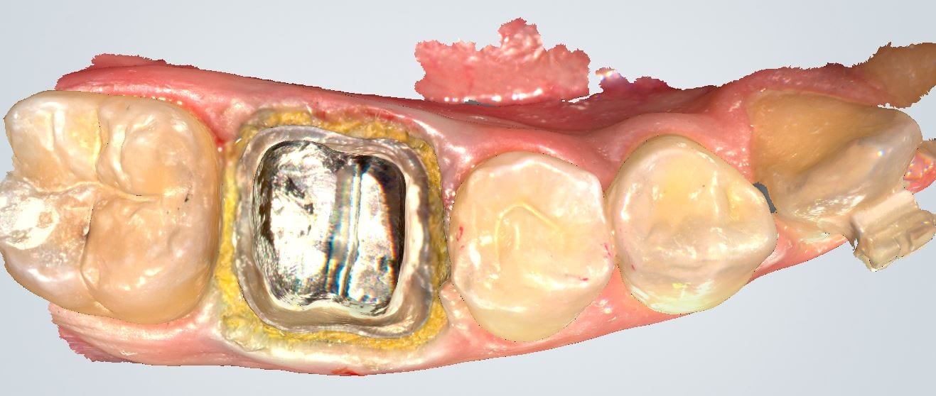
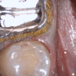
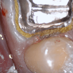
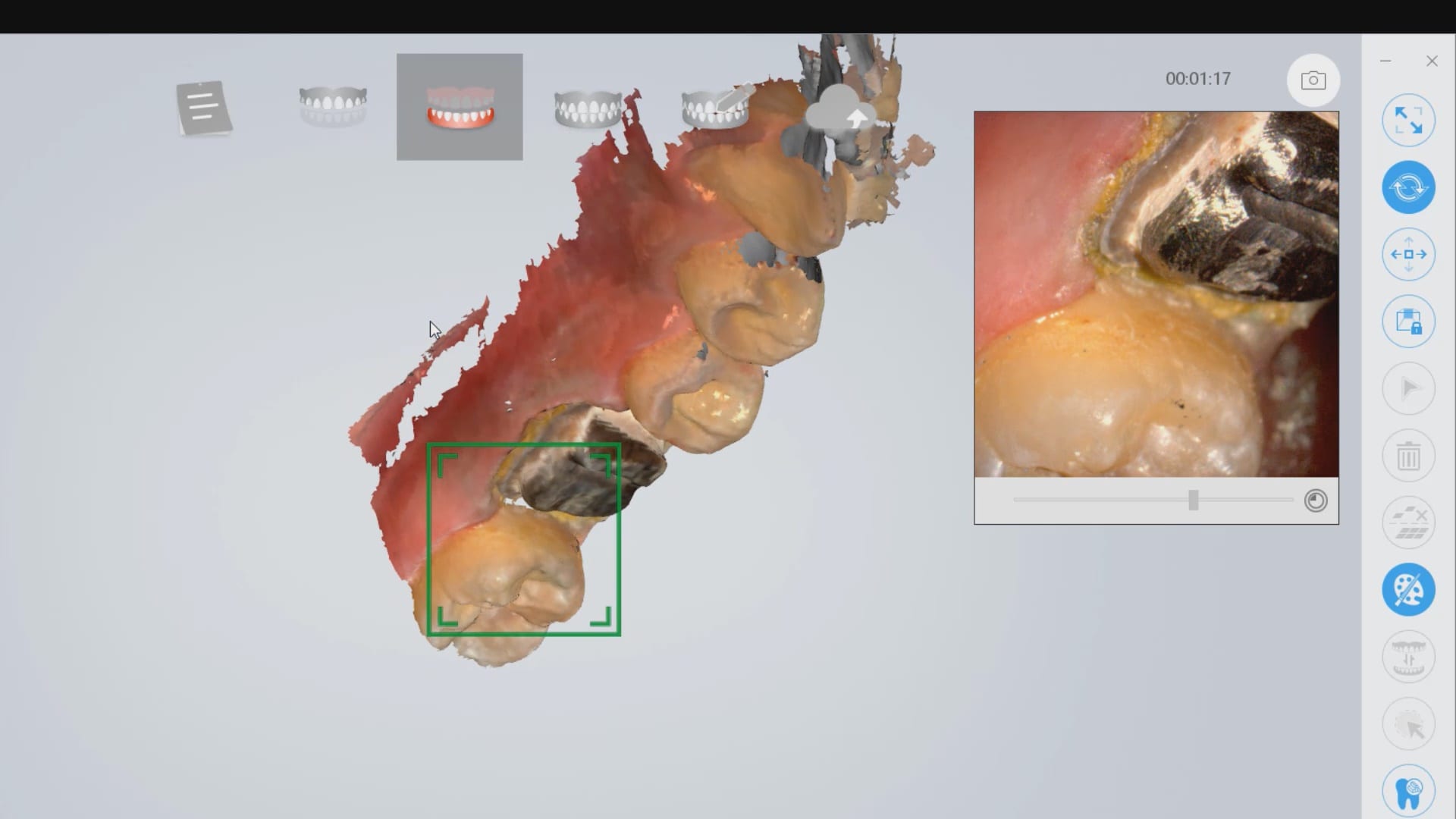
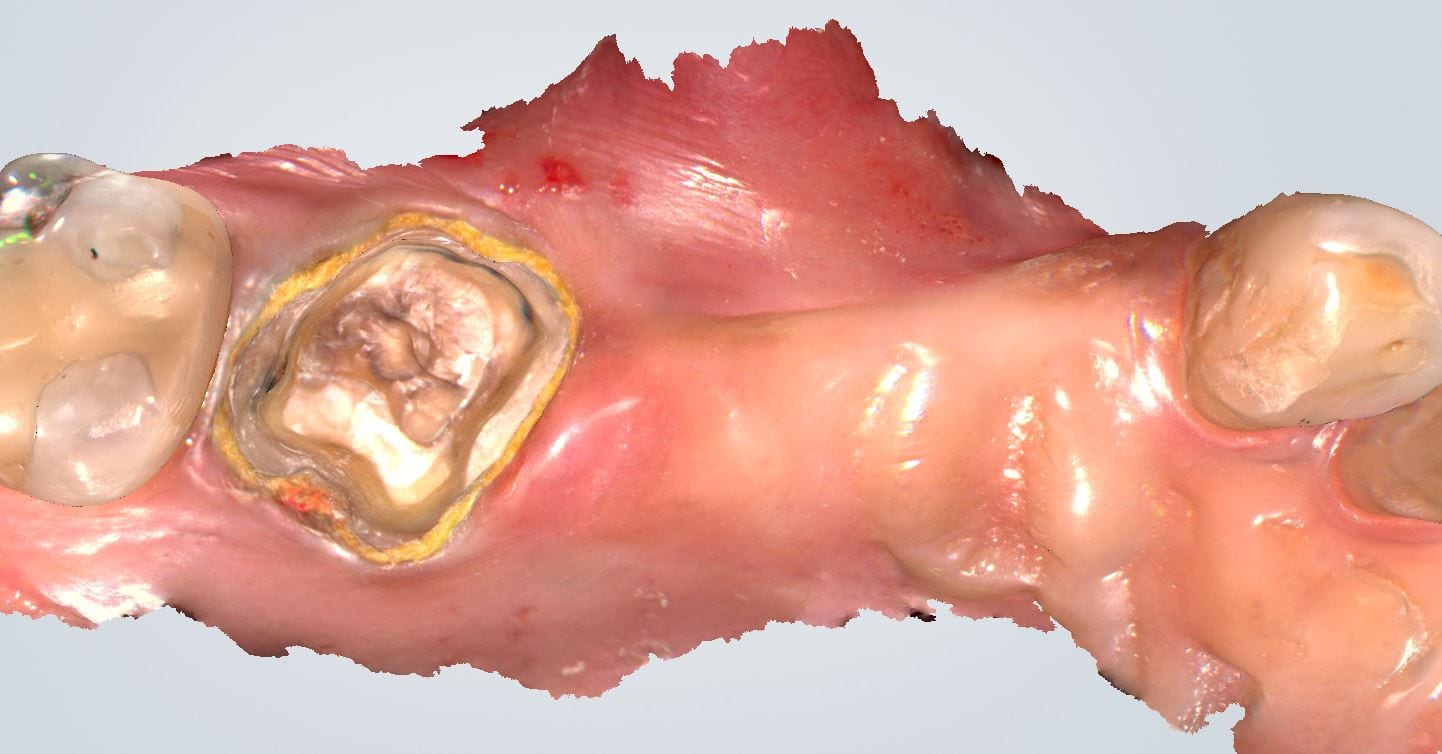
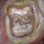
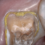
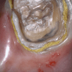
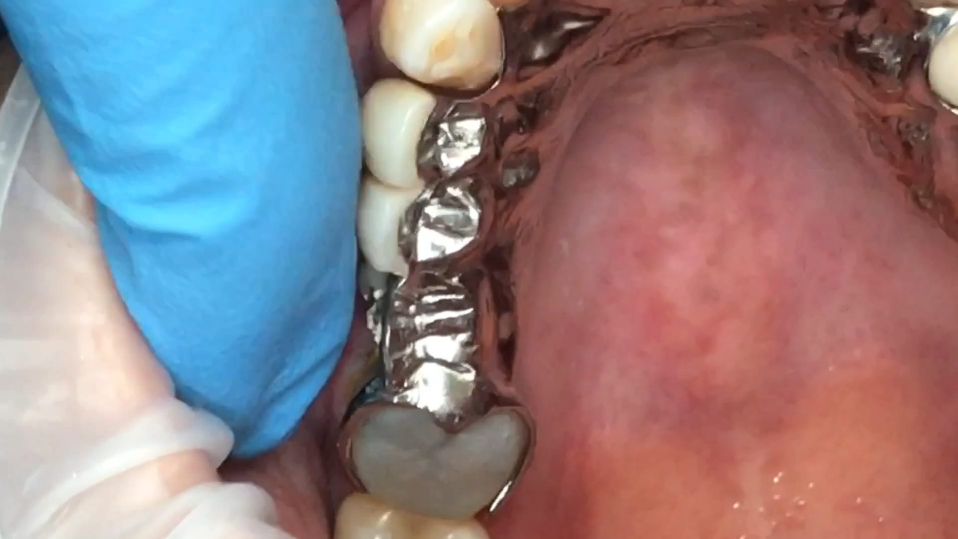
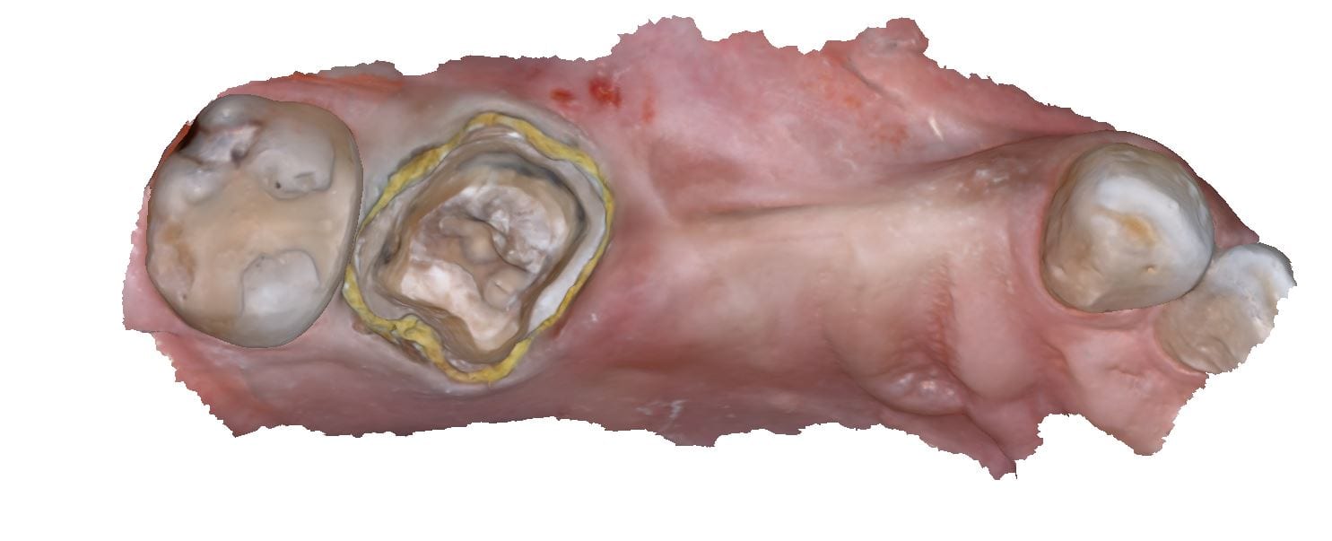
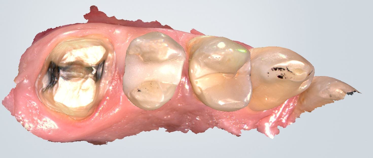
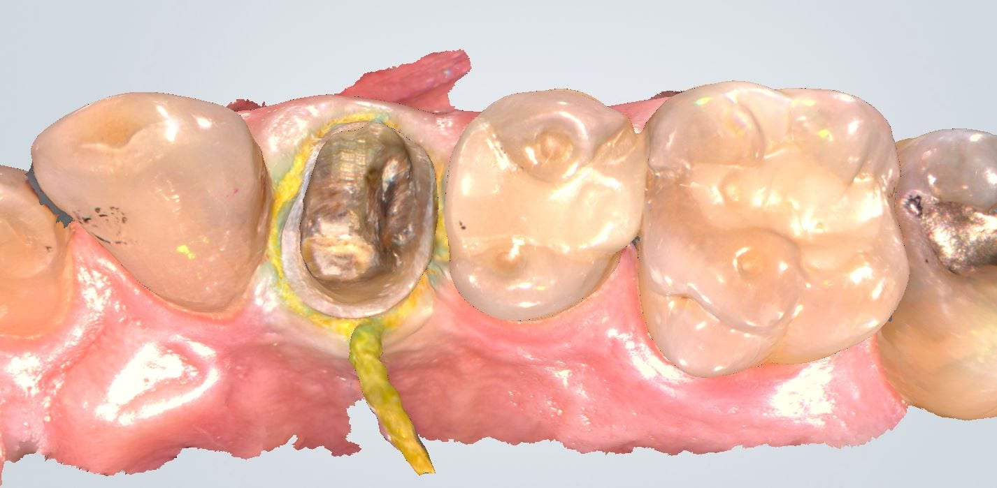
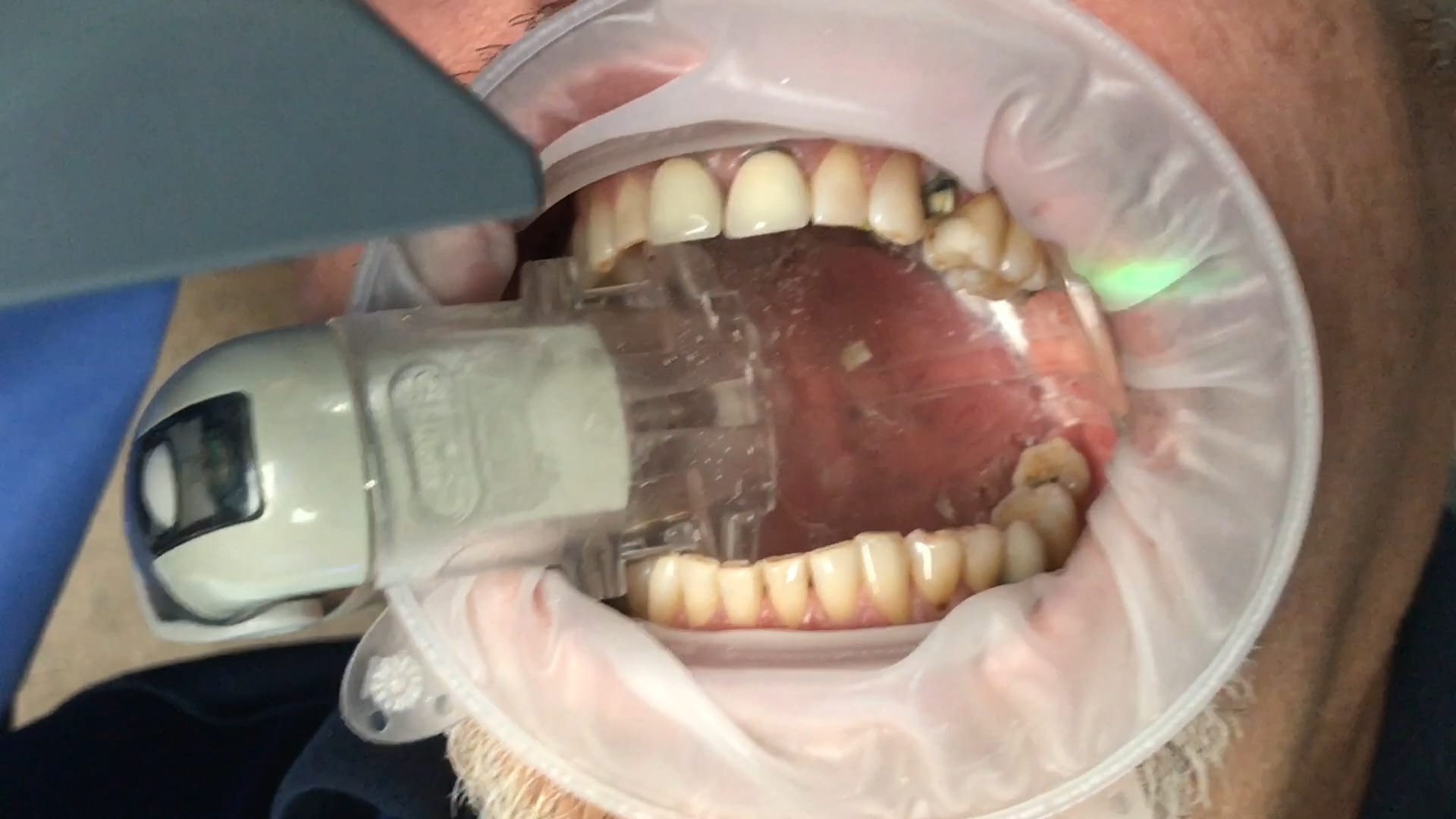
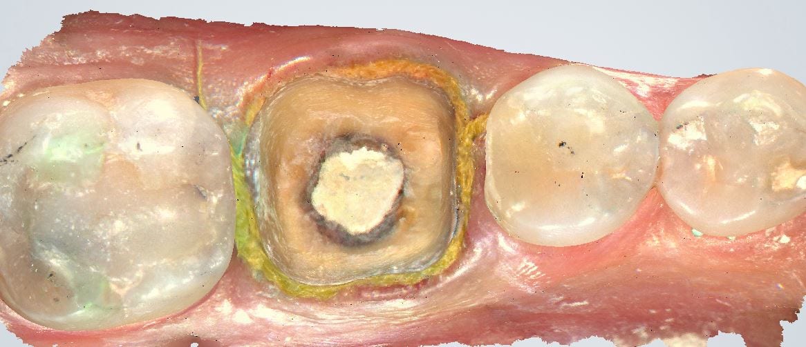
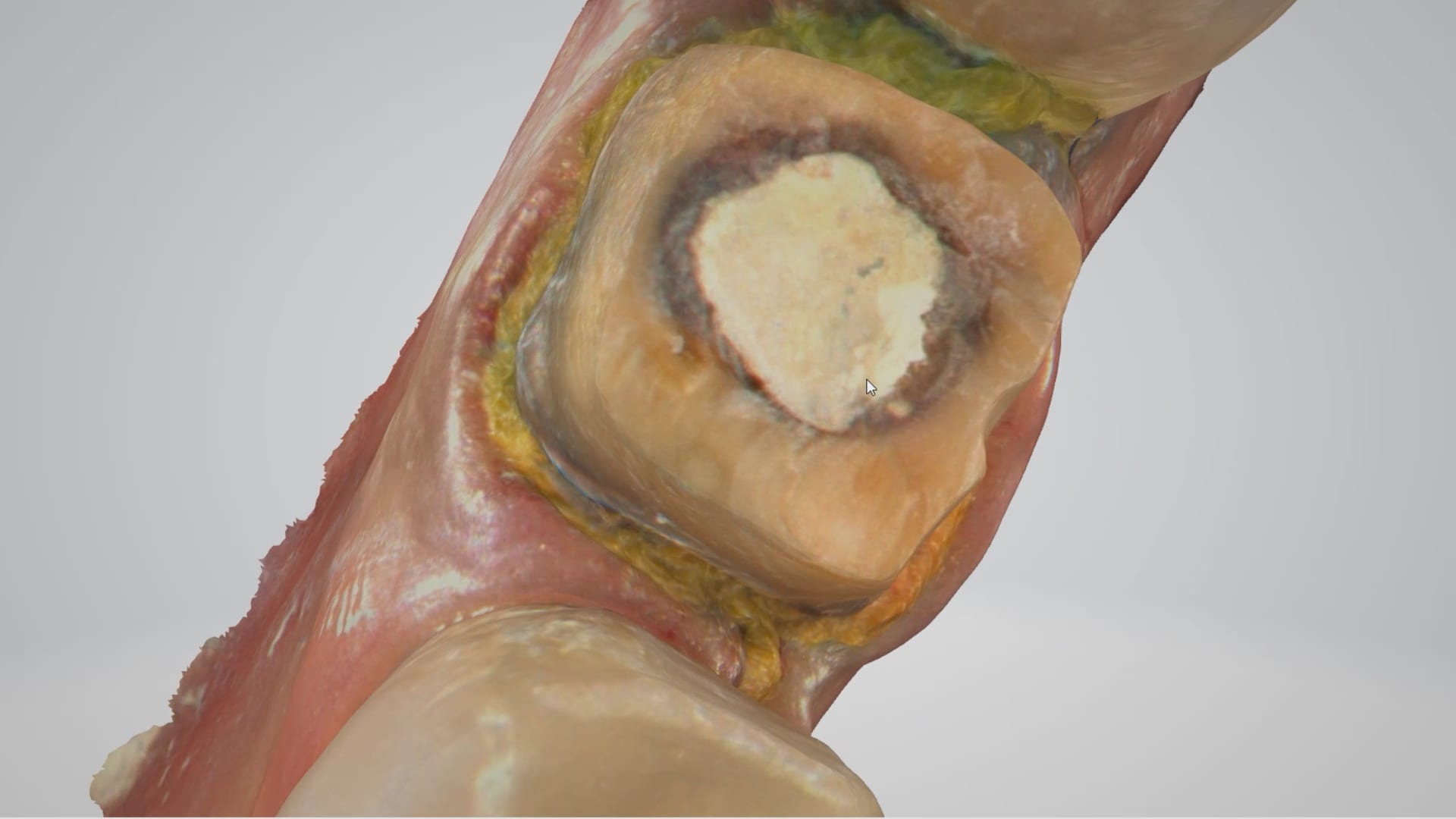
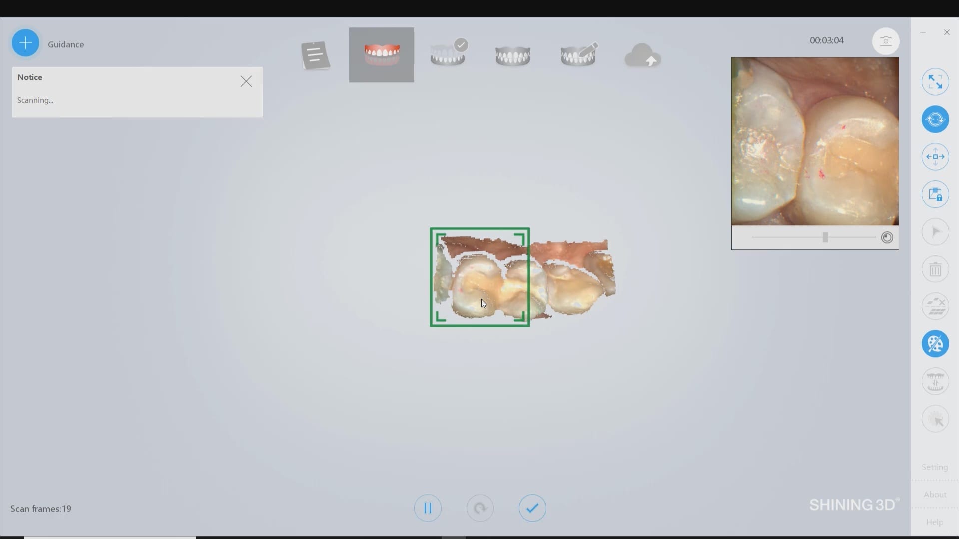
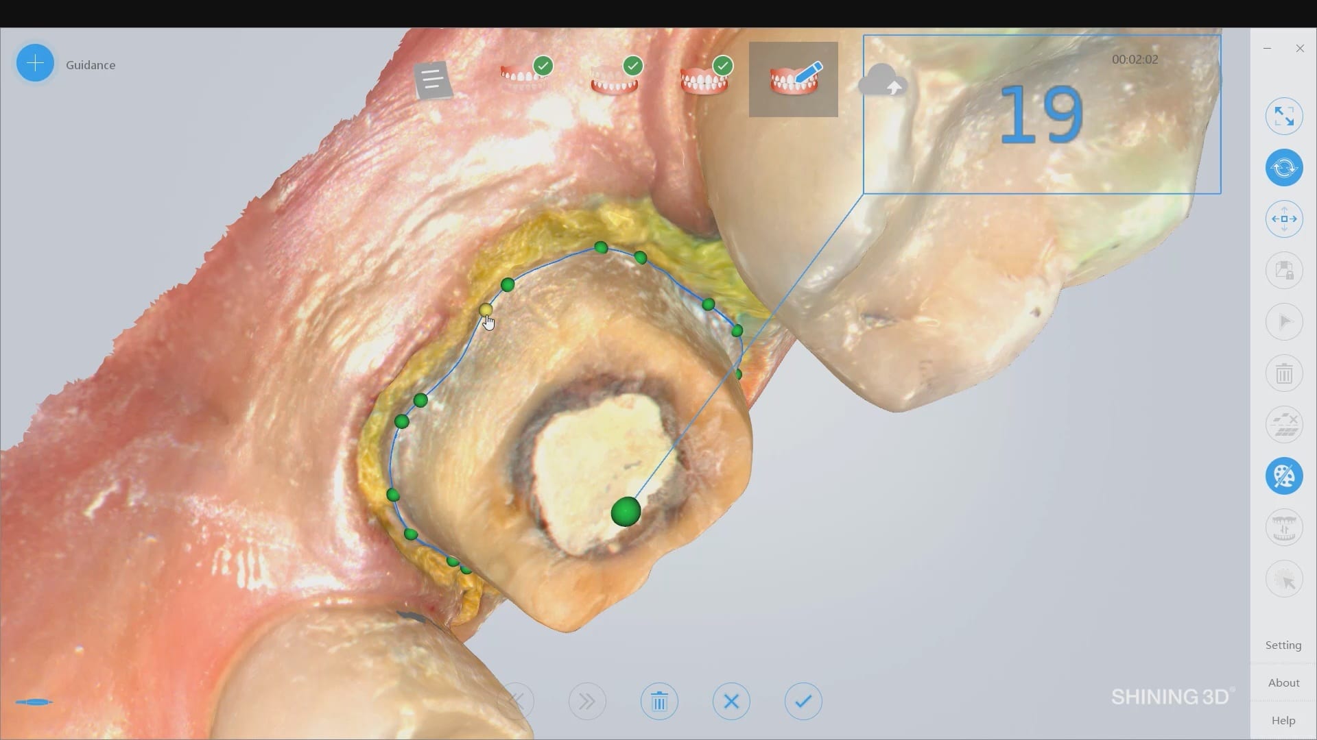
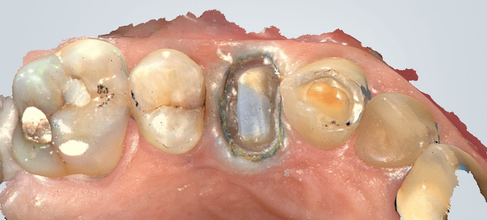
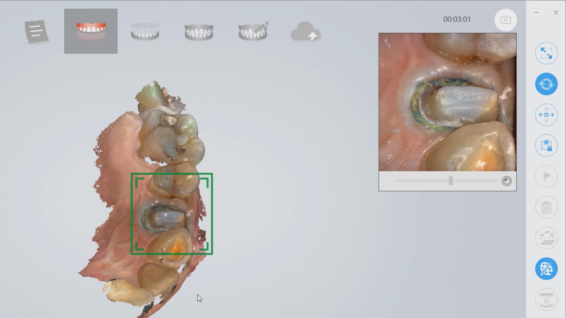
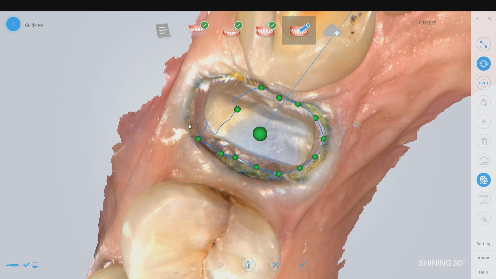
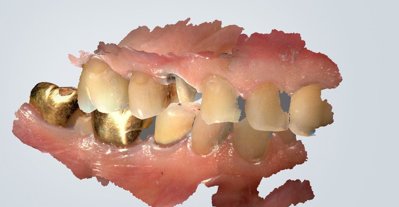
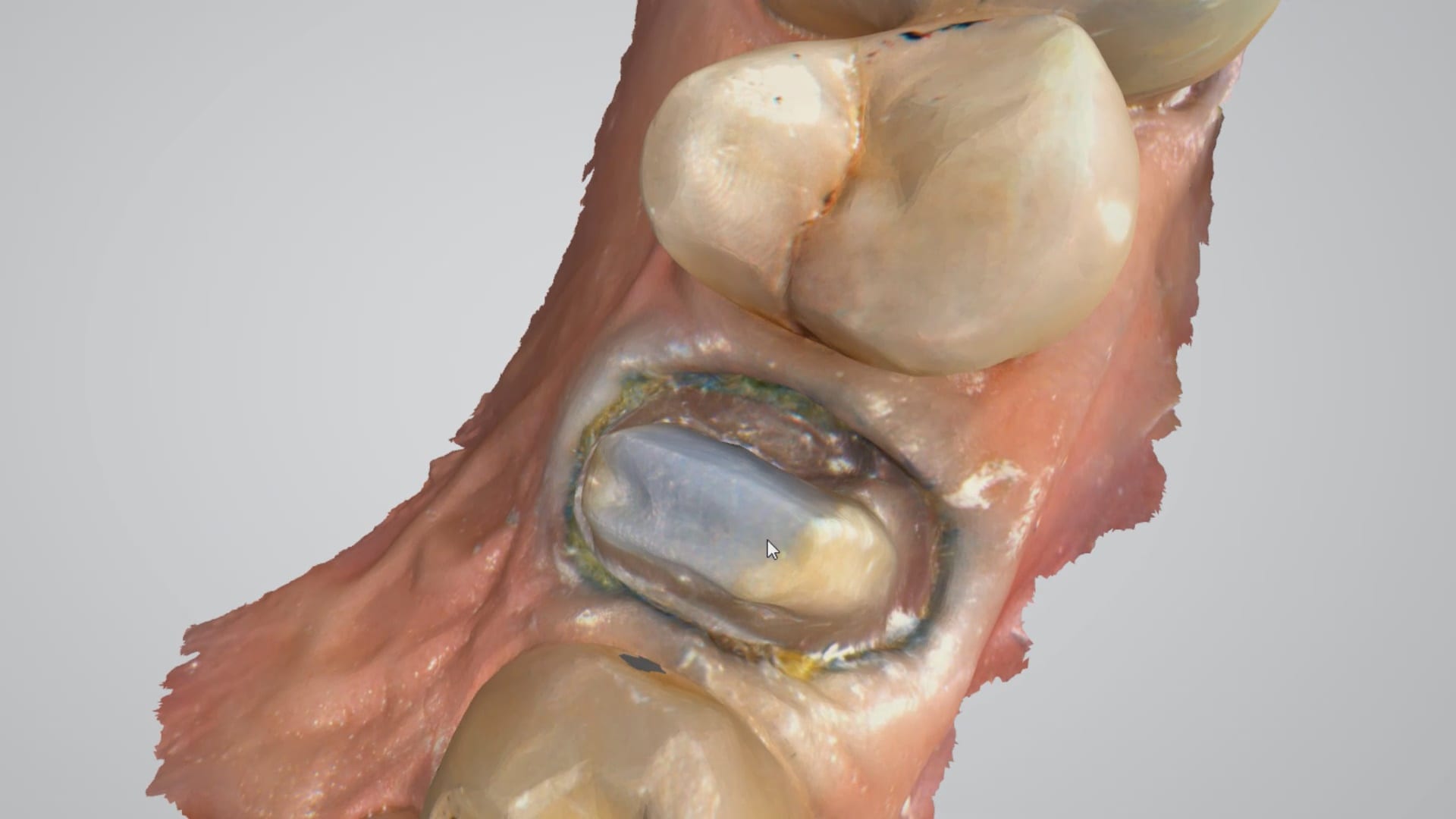
How Well does the Intraoral Scanner  Work on Soft and Hard Tissue?
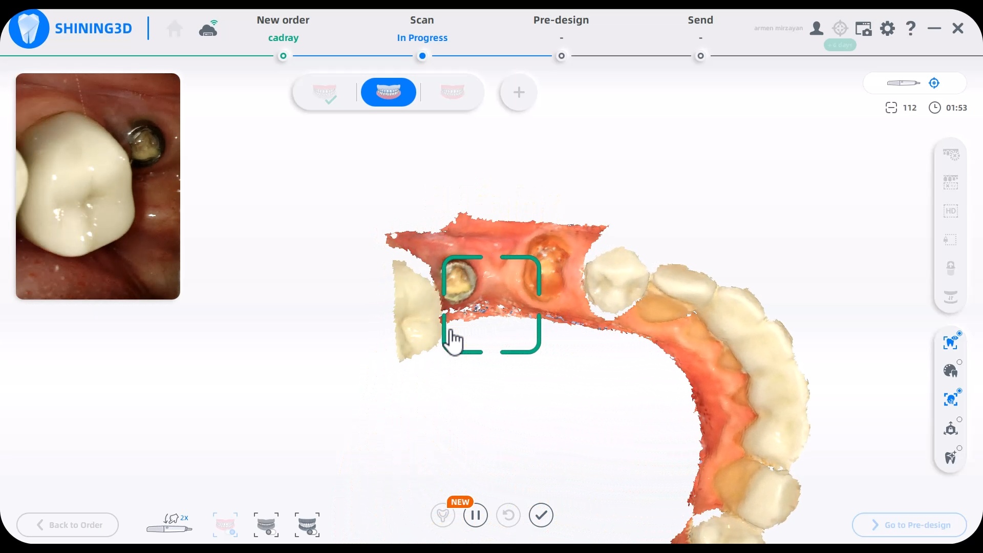
lots of people ask about the intra oral scanning experience with the shining elite device. please note that this is COMPLETELY different technology than the intraoral photogrammetry unit. it has performed on par with every other scanner i use, if not better. it has such a huge field of view it picks up a lot of data at once. It is prone to every issue that all ios have, ranging from double models to introducing errors over long edentulous and symmetric spans, etc… so scan path is still important.

video
Intra Oral Scanning Performance

play-rounded-fill play-rounded-outline play-sharp-fill play-sharp-outline
pause-sharp-outline pause-sharp-fill pause-rounded-outline pause-rounded-fill
00:00
03:12

Case 2

The first video in this series shows a quick overview of a multi unit case and the subsequent videos goes into more detail

video
Quick Overview

play-rounded-fill play-rounded-outline play-sharp-fill play-sharp-outline
pause-sharp-outline pause-sharp-fill pause-rounded-outline pause-rounded-fill
00:00
01:31
Item 1.Item 1.

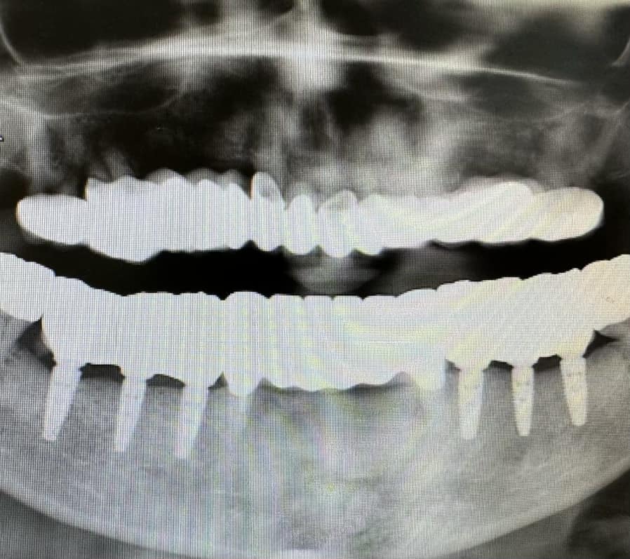
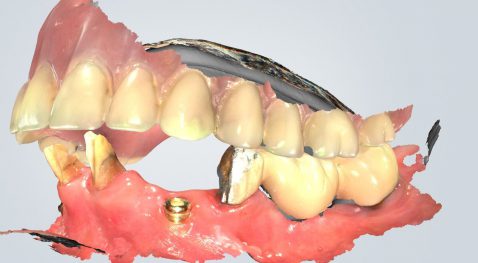
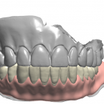
Radiograph of the delivered case


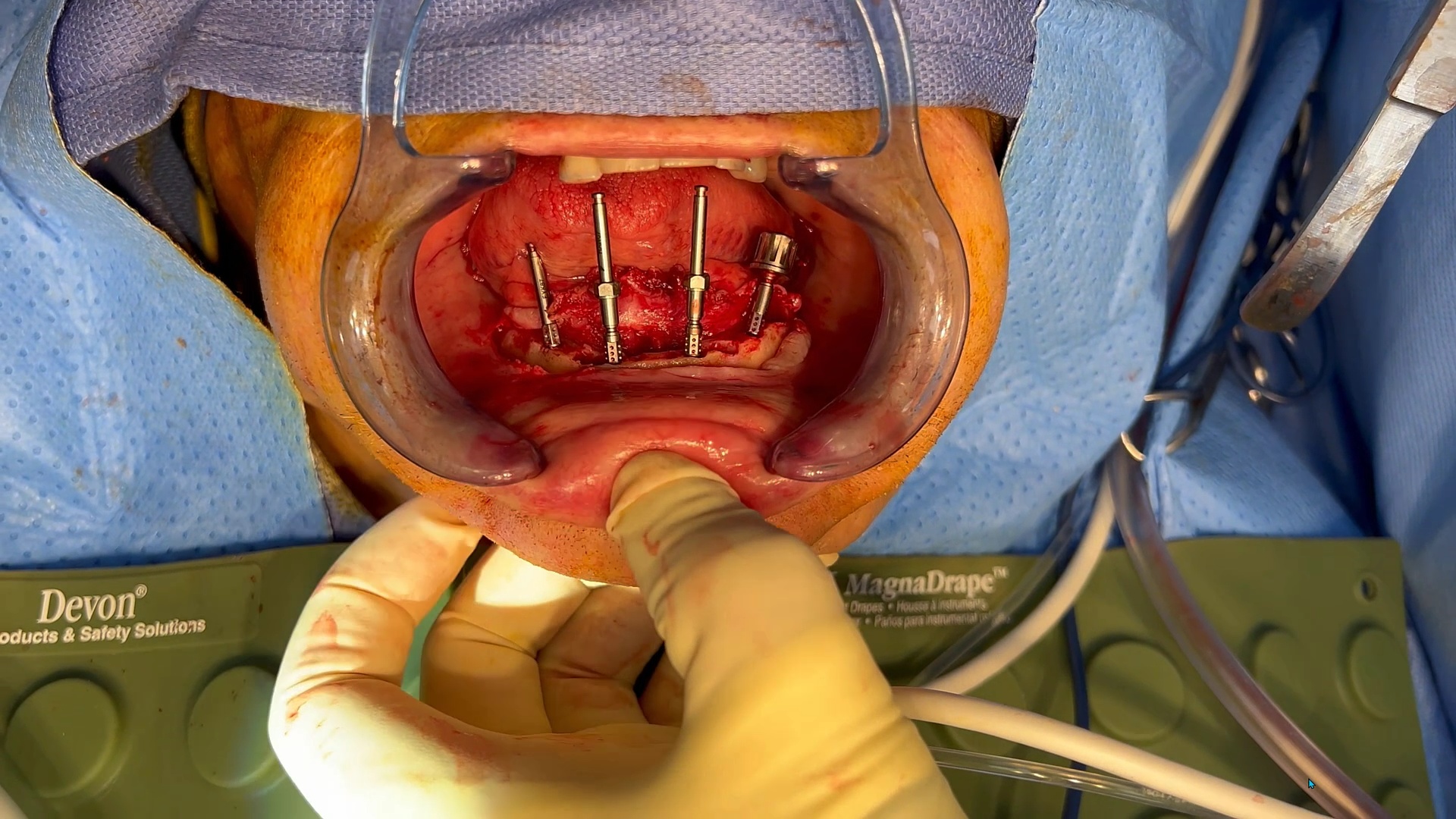
Case 3 – Managing and immediate case with bone reductio

This video shows how to manage immediate conversion cases and compares and IPG with an extra-oral Photogrammetry Machine

video

play-rounded-fill play-rounded-outline play-sharp-fill play-sharp-outline
pause-sharp-outline pause-sharp-fill pause-rounded-outline pause-rounded-fill
00:00
11:28

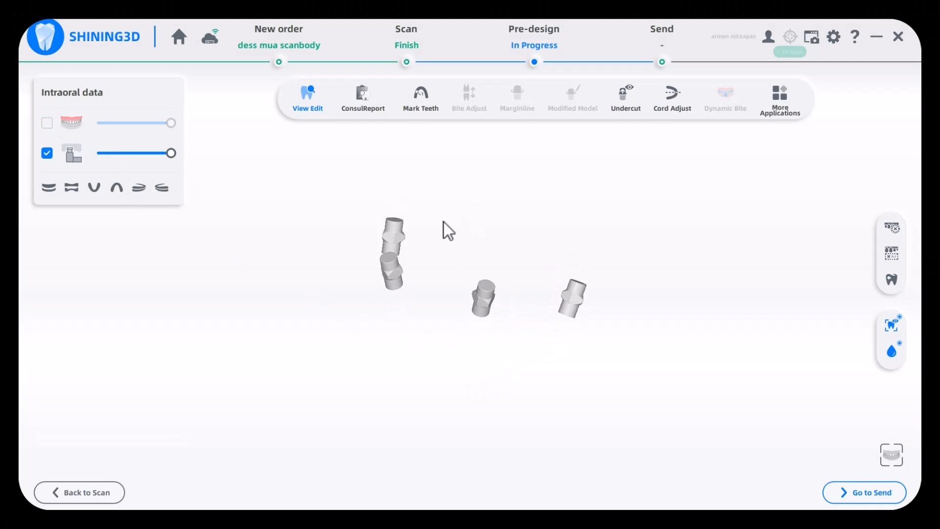
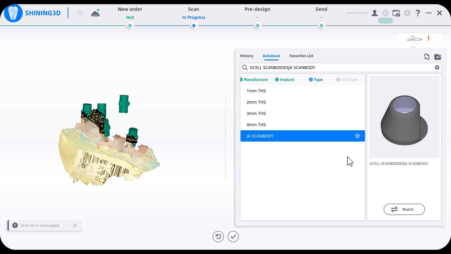
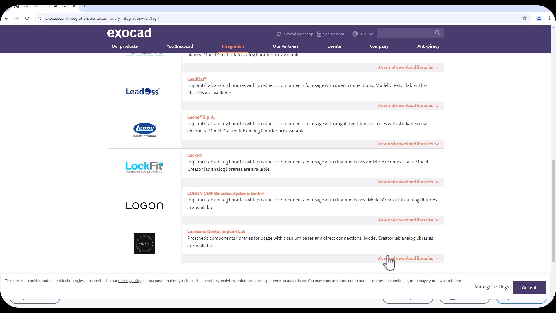
How to load libraries into Shining 3D Elite Intra-Oral Photogrammetry Software

This should be done by an experienced person.  Here we load a dess MUA library into the equation in the first video. In the second video we import Jonathan Abeneim’s Xcell libraries, followed by Danny Domingue Vortex library import

video

play-rounded-fill play-rounded-outline play-sharp-fill play-sharp-outline
pause-sharp-outline pause-sharp-fill pause-rounded-outline pause-rounded-fill
00:00
01:23
Item 1.Item 1.

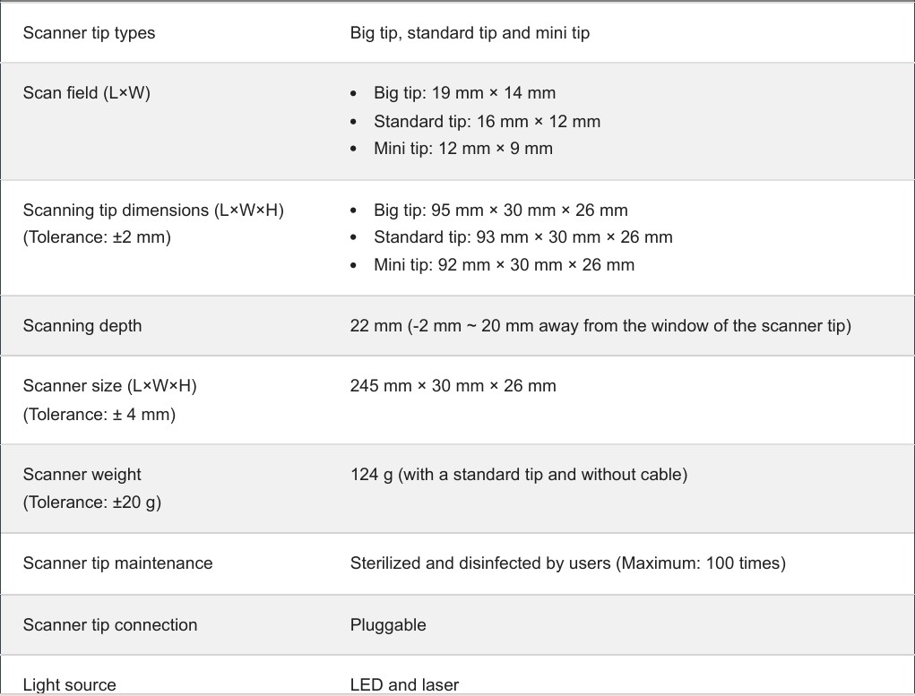
Scanner Tip Dimensions

Posted on Leave a comment

Single Click Launches Shining3D CAD Program

Shining3d has a projector and a single camera that takes photos of the surfaces it is scanning. at first, you may think this is a shortcoming, but in many ways, it dramatically reduces errors a new user can introduce.

in the early days of cerec, all you needed to do was capture the margins and the area above the height of contours of the adjacent teeth. This made the try-in and the contacts really easy as the design software just dropped straight a straight wall down to make contacts to. same concept here!

also, after you image and place margins, A SINGLE CLICK takes you to design software where you can finish the case and mill it right away.

video
shining3d single click launch into CAD software

play-rounded-fill play-rounded-outline play-sharp-fill play-sharp-outline
pause-sharp-outline pause-sharp-fill pause-rounded-outline pause-rounded-fill
00:00
00:00
Posted on Leave a comment

Virtual Extractions for Overdenture with Shining 3D

upper denture duplicated with shining 3d. only needed the occlusal and buccal surfaces as the case is for a lower overdenture over immediate extractions, on healed implants. went ahead and just duped the whole denture with the ios. very good results

took off lower temps and scanned lower jaw. remaining teeth were used for long term temp abutments while implants healed. digital extractions performed and right vertical dimension for digital prosthesis design

video
duplicating upper denture extra-orally

play-rounded-fill play-rounded-outline play-sharp-fill play-sharp-outline
pause-sharp-outline pause-sharp-fill pause-rounded-outline pause-rounded-fill
00:00
00:00

Full arch imaging of mandible in preparation of overdenture

video
lower arch scan for overdenture on healed abutments

play-rounded-fill play-rounded-outline play-sharp-fill play-sharp-outline
pause-sharp-outline pause-sharp-fill pause-rounded-outline pause-rounded-fill
00:00
00:00

buccal bite taken intra-orally to mount arches at proper vertical dimension.  you can see how the camera performs in this challenging case

video
intra-oral full arch bite with shining 3d

play-rounded-fill play-rounded-outline play-sharp-fill play-sharp-outline
pause-sharp-outline pause-sharp-fill pause-rounded-outline pause-rounded-fill
00:00
00:00

Digital extractions performed in preparation  for overdentures

video
virtual extractions for overdenture

play-rounded-fill play-rounded-outline play-sharp-fill play-sharp-outline
pause-sharp-outline pause-sharp-fill pause-rounded-outline pause-rounded-fill
00:00
00:00

Overdenture Design and Fabrication

Posted on Leave a comment

Full Arch Scan by The Shining 3D Aoralscan

In this video we demonstrate how the Aoralscan can complete a full arch scan utilizing the basic principles of digital impressioning.

Proper retraction, isolation, and scanning technique lead to high quality, error free models.


video
full arch scan by the shining 3D AoralScan by Nick Statly

play-rounded-fill play-rounded-outline play-sharp-fill play-sharp-outline
pause-sharp-outline pause-sharp-fill pause-rounded-outline pause-rounded-fill
00:00
00:00
Posted on Leave a comment

Lower First Molar Deep Margins Scanned by Shining AoralScan

In this video we demonstrate how the Aoralscan from Shinning 3D captures deep margins.

An old PFM crown warranted replacement due to open margins and recurrent decay. After the crown was removed, the margins were refined and the tissue was displaced with expasyl and retraction cord.

This video is deliberately captured slowly so a new user can appreciate how to hold the camera to capture the margins and the contacts of the adjacent teeth. You also have the opportunity to place your own margins and the path of draw before submit the case to the lab.

video
Shining 3D Aoralscan

play-rounded-fill play-rounded-outline play-sharp-fill play-sharp-outline
pause-sharp-outline pause-sharp-fill pause-rounded-outline pause-rounded-fill
00:00
00:00
Posted on Leave a comment

Shining 3D Aoralscan Sample Meshwork

This case was scanned by two different scanners. Once by the Medit i500 and another time with the Aoralscan. Both the meshes are included so you can compare the two. It was scanned to deliver a case that retrofits a partial denture

video
delivery of crown

play-rounded-fill play-rounded-outline play-sharp-fill play-sharp-outline
pause-sharp-outline pause-sharp-fill pause-rounded-outline pause-rounded-fill
00:00
00:00

Download the Medit Scan

Download the case from Shining


Posted on Leave a comment

Upper Right First Molar Scanned with the Shining 3D Intra-Oral Scanner for Beginners

In this case presentation, we introduce the Shining Aoralscan for beginner doctors who want to get into digital impressions.  Proper isolation and retraction is a key element of scanning. In this demonstration we showcase how a new user can easily control the operating field to deliver high quality restorations

video
imaging with shining 3D AOralscan

play-rounded-fill play-rounded-outline play-sharp-fill play-sharp-outline
pause-sharp-outline pause-sharp-fill pause-rounded-outline pause-rounded-fill
00:00
00:00
Posted on Leave a comment

Upper Left Premolar with Shining 3D

Proper isolation and retraction is key for all intra-oral scanners.  In this video, you can appreciate how the upper and lower arches were isolated, the margins were exposed with retraction cord and imaged with the shining 3d aoralscan

video
Isolation with isolite and optragate for imaging with Shining 3D

play-rounded-fill play-rounded-outline play-sharp-fill play-sharp-outline
pause-sharp-outline pause-sharp-fill pause-rounded-outline pause-rounded-fill
00:00
00:00

play-rounded-fill play-rounded-outline play-sharp-fill play-sharp-outline
pause-sharp-outline pause-sharp-fill pause-rounded-outline pause-rounded-fill
00:00
00:00

Download the obj file


Posted on Leave a comment

Shining 3D AoralScan Margins

video
preview of margins captured by Shining 3D Aoralscan
html5: Video file not found

pause-sharp-outline pause-sharp-fill pause-rounded-outline pause-rounded-fill
video
case set up and imaging of upper and lower arches and buccal bite

play-rounded-fill play-rounded-outline play-sharp-fill play-sharp-outline
pause-sharp-outline pause-sharp-fill pause-rounded-outline pause-rounded-fill
00:00
00:00
video
labeling tooth and marking margins

play-rounded-fill play-rounded-outline play-sharp-fill play-sharp-outline
pause-sharp-outline pause-sharp-fill pause-rounded-outline pause-rounded-fill
00:00
00:00

Click to Download the Lower Arch Model OBJ file

Posted on Leave a comment

Our First Shining 3D Aoralscan Case- A Preview

At CAD-Ray, we are big proponents of open architecture and the doctors control the flow of their patients’ digital data, whether it is a CT scan or an digital impression system.  We have put the Aoralscan through a battery of tests. For single unit, it delivers on quality that equals any other scanner on the market.  We were particularly impressed with this deep margin and how well the graphics could differentiate tissue from tooth structure on the distal of the prep

image upper and lower arch with shining 3d aoralscan

play-rounded-fill play-rounded-outline play-sharp-fill play-sharp-outline
pause-sharp-outline pause-sharp-fill pause-rounded-outline pause-rounded-fill
00:00
00:00
Item 1.Item 1.

click to download the OBJ file of this case (note: we only scanned to pick up data for the margins)

video
check out how well the shining 3d aoralscan picks up deep margins

play-rounded-fill play-rounded-outline play-sharp-fill play-sharp-outline
pause-sharp-outline pause-sharp-fill pause-rounded-outline pause-rounded-fill
00:00
00:00